Download
1 / 1
10 likes | 172 Views
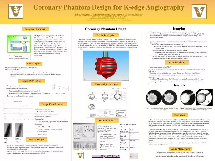
Overview of MX200. Device Description. Market Analysis. Subtraction Method. Social Impact. Hypersensitivity reaction to iodine (3-12% patients) Increase patient safety: reduce the amount of potent contrast agents used during angiography

E N D
Overview of MX200 Device Description Market Analysis Subtraction Method Social Impact • Hypersensitivity reaction to iodine (3-12% patients) • Increase patient safety: • reduce the amount of potent contrast agents used during angiography • decrease the dose of ionizing radiation to the patient by employing K-edge Imaging techniques • Product is a reusable coronary phantom specific for imaging research for the MX200. • The determination of the optimal angle for stereoscopic imaging as a function of distance will benefit the radiological community. • Optimal cost to performance ratio with an associated manufacturing cost of $20 for materials testing, while the current industry manufacturing cost is ~$5000. • Design a coronary phantom • Test contrast agent concentrations • Current clinical dilution with saline solution is 1:16 • Using monochromatic plain films it is possible to see 1:256 dilutions • Determine optimal angle for stereo imaging • Project Deliverables 3” θ 30” Figure 2. Initial images taken with an empty phantom (left) and with saline (right). Figure 3. Images taken with 1:256 dilution with the subtraction method (left) and differential method (right). Design Considerations • Radio translucent at 35keV • K-edge of Iodine is 33.2KeV • Flexible material for a pulsatile model • Fluid perfusion capabilities • Phantom Size • 7.5cm diameter Approximate size of canine heart • Vasculature • Size Range 5 - 1 mm • Coronary arteries represented on surface • Curved channels • Hollow vessels Material Testing Acknowledgements Phantom Specifications Conclusion Coronary PhantomDesign for K-edge Angiography John Jorgensen1, Sarah Pachtman1, Punam Patel1,Marcus Spallek1 Advisors: Paul King, Ph.D.1;Frank E. Carroll, M.D.2 1 Department of Biomedical Engineering, Vanderbilt University, Nashville, TN 2Vanderbilt University Medical Center Department of Radiology and Radiological Sciences Imaging Coronary Phantom Design • The phantom was not constructed with the material that was specified. Due to the specifications of the printing machine used to manufacture the prototype, Z-Corp’s ZP-102 plaster-based powder was used to create the prototype. Images were shot using a semi-monochromatic method: • Images were taken with two polychromatic tubes running at 50KVP, having different effective X-ray energies: • Molly tube (Molybdenum) with an aluminum filter. • gives an X-ray spectrum with a peak around 18KeV provided our “below the K-edge of Iodine” shots. • Tungsten tube with an aluminum filter running at 50KVP. • has a peak X-ray output in the range of 50 KeV (values rounded off in data analysis as 50KeV since it is still simply polychromatic) • Results were imaged on MAR345 digital detector running in 100µ resolution mode. They were then downloaded to a PC and processed using ImageJ. MXISystems, Inc. in conjunction with Vanderbilt University and the Office of Naval Research has developed a laser synchrotron (Inverse Compton scattering) X-ray source for the energy region from 10 keV to 50 keV. It is capable of producing X-ray fluences sufficiently high that a single, 9-ps pulse produces a complete medical X-ray image. The device is a tunable, monochromatic x-ray (MX200) to be used in medical imaging. Current research focuses on the application for intravenous coronary angiography. The coronary phantom consists of a hollow chamber, with a 5 mm diameter tube for saline pump attachment. The outer surface is comprised of interconnected tubes. The three vertical tubes have internal diameter of 1 mm. The horizontal tubes range in diameter from 2 to 5 mm. Two openings are directly connected to the ring grid and allow for fluid filing and emptying. The tubes do not open into the chamber. The device is one solid unit designed to be filled with fluids for imaging with the MX200. Figure 1. Diagram of MX200’s laser paths and major elements. http://www.mxisystems.com/mx200.html • Images were taken at 18 and 50KeV • A portion of incident X-rays was selected and the exposure was summed in the same area of both images. • The images were normalized to each other so that the area of selection of each image contained the same exposure by multiplying the lesser exposed by the ratio of the two selections. • Using ImageJ Image Calculator the “difference” between the high energy and the low energy was created, making the areas that might have increased absorption due to k-edge effects more pronounced. Results Tan θ = 3/30 = 0.10 θ ~ 6° 3-D images of the empty phantom indicated that one of the walls separating the arteries from the main chamber was damaged. It was noted that when air was blown into the arterial portion of the phantom the air leaked out of all ports, indicating the presence of a hole in the wall between the inside chamber and the surface arteries. The phantom was filled with a small amount of saline and a leak was detected. The images were taken with an iodine dilution of 1:256. A balloon filled with water was inserted in the center chamber while the contrast solution was filled in the arteries (image on left). Note the balloon and water appear very opaque and would require high dose exposures to image through the phantom. The 1:256 dilution we were able to image is still significantly lower than the dose of about 1:16 given to a CT patient. The image on the right was edited using a differential method. This method relies on logic function and provided a clear image of the contrast agent over the subtraction method. The 3-D movie shows the phantom with rotations of 3, 6, 9, 12 degrees. The 6° is found to be the optimal stereo angle for imaging. • Several samples of 2-component polyurethane resins tested for radio opaque appearance at 35 keV. • Smooth-On Reoflex™ 20 liquid rubber chosen as ideal material because of translucent appearance on detector. • High grayscale value indicates degree of x-ray absorbance and translucency. Finger imaged at 26 keV Finger imaged at 19 keV This project was made possible through the help of the following contributors: Scott Degenhardt; Robert Traeger; Dr. Frank Carroll; Philip Davis; Z-Corporation