Download

1 / 112

E N D
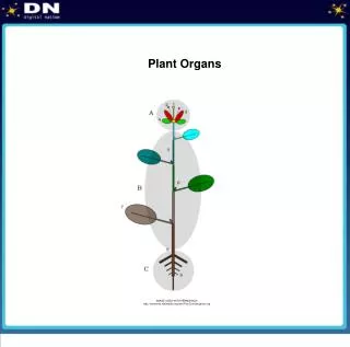
By Jessica Lewis Organs
Functions: • The Skeletal System serves many important functions. It provides the shape and form for our bodies. It also supports, protects allows our bodies to move, allows our bodies to function, produces blood for the body, and also store minerals.
Functions: cont. • Many vital organs are protected by the skeletal system. One example is the brain, it is protected by the skull while the lungs and heart are protected by the ribcage. Our bodies function because there is an interaction between our muscles and skeletal system. Muscles are connected to the bone by tendons. Now, bones are connected to each other by ligaments . Muscles which cause movement of a joint are connected to two different bones and contract to pull them together. An example would be the contraction of the biceps and a relaxation of the triceps. Blood cells are produced by the marrow located in some bones. Around 2.6 million red blood cells are produced every second by the bone marrow to replace the ones that have become worn out and destroyed by the liver. Bones serve as a storage area for minerals such as calcium and phosphorus. When an excess is present in the blood, buildup will occur within the bones. When the supply of these minerals that is with in the blood is low, it is withdrawn from the bones and is replenished.
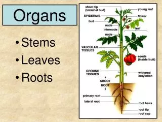
Divisions of the Skeleton: Axial: Appendicular: • This consists of bones that form an axis of the body and protect the organs of the head, neck and trunk. It also supports these organs. These bones protect the: • The Skull • The Sternum • The Ribs • The Vertebral Column • This skeleton is composed of bones that anchor the appendages to the axial skeleton. The organs that are involved with this are: • The Upper Extremities • The Lower Extremities • The Shoulder Girdle • The Pelvic Girdle
The Skull: • Your skull provides the framework for most of your sensory organs, such as eyes, ears, tongue, nose, and some skin. Your skull is made up of 22 cranial or facial bones, plus the three in each ear. As a baby you have more. Most are fixed joints separated by cartilage as a baby, but fuse together a later as you grow. Once fused, they are locked together, forming an immovable joints, called a suture-sound.
The Backbone: • The backbone, or vertebral or spinal column, though called a "bone", is really a flexible structure made of 26 bones. As a baby, you have 33 vertebrae, or back bones, but the lower four fuse to form the coccyx-sound, and the next lower five fuse to become the sacrum. The backbone serves several important functions itself. It provides structure from which all other upper body structures branch, and it protects the spinal nerve, which is the "highway" that all the information your brain sends to your body travels. The backbone is approximately 70 cm, long and is separated into five regions.
The Thorax: • The thorax is basically your chest, comprising your breast bone and ribs. Your breast bone, or sternum-sound, is around 6 inches (15 cm) tall, spanning about half the length of your ribs. The twelve ribs form the cage of your chest. One of the primary necessities of the ribs is to protect your heart and lungs. Your top seven ribs are called true ribs because they connected to the sternum. The next four ribs are called false ribs because they attach to the sternum so indirectly if at all. If they do not connect to the sternum, they do connect to upper cartilage for support. The last two ribs are called floating ribs because they do not connect to anywhere.
Types of Bone: Long Bones: Short Bones: • The long bones are longer then they are wide and they work as a lever. The bones of the upper extremities are where these bones could be found. The upper extremities are the humerus, tibia, femur, ulna, metacarpals ect. • Short bones are a cubed like and short. You can find them in wrists and ankles.
Types of Bone: cont. Flat Bones: Irregular Bones: • Flat bones have more of a broad service and are used to protect organs and attachments of muscles. For example the rib cage, cranial bones, and the bones of shoulder girdle. • Irregular bones are the bones that do not fall into any of the other categories. They all vary in size and shape and some can be found in the skull.
Bone Composition: • Bones are composed of tissue that may take one of two forms. Compact or spongy. Compact bone is very hard and dense and forms sort of a protective shield around the exterior of the bone.
Bone Composition: cont. • Spongy bone is inside of the bone and has a lot of holes that reside within it. You can find spongy bone in most all bones. The bone tissue is made up of several kinds of bone cells that are imbedded in sort of a web or inorganic salts like calcium. This gives the bon e strength and collagenous fibers allow the bone to be fexible.
Function: • The main role of the muscular system is to provide movement. Muscles work in pairs to move limbs and provide the organism with mobility. Muscles also control the movement of materials through some organs, such as the stomach and intestine, and the heart and circulatory system.
Cardiac Muscle: • The cardiac muscles is the muscle of the heart itself. The cardiac muscle is the tissue that makes up the wall of the heart called the mydocardium. Also like the skeletal muscles, the cardiac muscle is striated and contracts through the sliding filament method. However it is different from other types of muscles because it forms branching fibers. Unlike the skeletal muscles, the cardiac muscle is attached together instead of been attach to a bone.
Skeletal Muscle: • The skeletal muscle makes up about 40 % of an adults body weight. It has stripe-like markings, or striations. The skeletal muscles is composed of long muscle fibers. Each of these muscles fiber is a cell which contains several nuclei. The nervous system controls the contraction of the muscle. Many of the skeletal muscle contractions are automatic. However we still can control the action of the skeletal muscle. And it is because of this reason that the skeletal muscle is also called voluntary muscle.
Smooth Muscle: • Much of our internal organs is made up of smooth muscles. They are found in the urinary bladder, gallbladder, arteries, and veins. Also the digestive tract is made up of smooth muscle as well. The smooth muscles are controlled by the nervous system and hormones. We cannot consciously control the smooth muscle that is why they are often called involuntary muscles.
Major Skeletal Muscles: Facial: Neck: • In the facial are one finds all the muscles which move the face. Orbicularisoculi-sound are the two muscles that move the eye are. Frontalis-sound and Temporalis-sound are the two muscles which move the forehead and sides of your head. Zygomaticus-sound ands Masseter-sound are the two muscle that work in conjunction to move your jaw and upper lip area. Orbicularisoris-sound is the muscle which moves your lips • The neck area is almost entirely moved by the sternohyoid-sound and Sternocleidomastoid-sound. These muscles allow the neck to move your head left and right. They work with the platysma muscle to control how far you can move your head left and right. What allows your head to move up and down is the trapeziums'-sound. The trapezius is so large that it extend down to the shoulder and thorax area. The trapezius is a good example of how some muscles are named by their shape. the trapeziums looks just like a trapezoid.
Major Skeletal Muscles:cont. Shoulder: Arm: • A group of muscles all work together to move the whole shoulder area. This group takes into account the trapezius-sound, deltoid-sound, infraspinatus-sound, teres major-sound, and the rhomboid major-sound. The rhomboid major is called so because its shaped like the geometric shape of a rhombus. Along with the help of the ball and socket joint-hyperlink in your shoulder, these group of muscles allow your arm to throw a softball, pick things over your head, and give your arms a good strecth early in the morning. • Most known amongst teenage weight lifters is the arm area. The famous bicep brachii-sound is the muscle that allows you to bring your forearm close to your body and form a huge ball of muscle which catches a lot of attention amongst weight lifters. The tricep brachii-sound and brachialis-sound are the two other muscles located in the arm region.
Major Skeletal Muscles:cont. Forearm: Thorax: • A majority of the muscle in the forearm help control a part of the arm. Amongst these is the Berachiodialis major-sound, palmaris longus-sound, and Flexor carpi radialis-sound. The name of the flexor carpi radialis is a good example of how muscles are named by their function and location. This muscle is named carpi because of the bones that it helps move, the carples. Also, the name of radialis is made by the bone that its attached to, the radius. • The thorax is the set of muscles which carrying your head, arms, stomach, and any other upper body areas. These muscles are the trapezius-sound and latissimus dorsi-sound. Usually, the majority of the muscles of the thorax can be damaged easily is one dose not stretch before exercise, or lifts a heave load.
Major Skeletal Muscles:cont. Abdomen: Hip: • The abdominal area consists of the muscles that allows you to bend down and move your waist from side to side. The interanal oblique-sound and external oblique-sound are the muscles that move your body from left to right. The Transversus abdominus-sound and Rectus abdominus-sound, along with the trapezius-sound an latissimus dorsi-sound allow you to bend down and grab objects. • Only two muscles make up the hip area. These are the gluteus medius-sound and gluteus maximus-sound. Probably the laziest muscles in the whole system the gluteus set of muscles are used only to sit down on.
Major Skeletal Muscles:cont. Pelvis/ Thigh: Leg: • An overlapping of muscles is what makes this area so firm. The pelvis area is usually referred to as the upper part of the leg. Muscles like the pectineus-sound and illiopsoas-sound , which help support the upper leg area are known as pelvic muscles. Thigh muscles are very rich in capillaries and support the whole body. The upper thigh muscles are abductor longus-sound, Gracilis-sound, Sartorius-sound, and Tensor fasciae latea. The lower thigh muscles are rectus femoris-sound, vastus lateralis-sound and medialis-sound. Located in the back of your leg are the hamstrings-sound. These muscles help you run, jump, and walk! • Helping the thigh region support the body is the Leg region. These muscles like the Gastrocnemius-sound, soleus-sound, porenius longus-sound, and Tibialis anterior-sound absorb the impact when one walks and runs. they also give beter cordination for moving. the thigh region trust the body forward while the leg region coordinates where it should be thrusted and where it should stand.
Function: • The main role of the circulatory system is to transport nutrients, gases (such as oxygen and CO2), hormones and wastes through the body.
Parts of The Circulatory System • The Heart • The Blood • The Blood Cells
The Heart: • The heart beats about 3 BILLION times during an average lifetime. It is a muscle about the size of your fist. The heart is located in the center of your chest slightly to the left. It's job is to pump your blood and keep the blood moving throughout your body.
The Blood: • The blood is an amazing substance that is constantly flowing through our bodies. • Your blood is pumped by your heart. • Your blood travels through thousands of miles of blood vessels right within your own body. • Your blood carries nutrients, water, oxygen and waste products to and from your body cells. • A young person has about a gallon of blood. An adult has about 5 quarts. • Your blood is not just a red liquid but rather is made up of liquids, solids and small amounts of oxygen and carbon dioxide.
The Blood Cells: Red Blood Cells: White Blood Cells: • They are responsible for carrying oxygen and carbon dioxide. Red Blood Cells pick up oxygen in the lungs and transport it to all the body cells. After delivering the oxygen to the cells it gathers up the carbon dioxide and transports it back to the lungs where it is removed. • White Blood Cells help the body fight off germs. White Blood Cells attack and destroy germs when they enter the body. When you have an infection your body will produce more White Blood Cells to help fight an infection.
The Blood Cells: cont. Platelets: • Platelets are blood cells that help stop bleeding. When we cut ourselves we have broken a blood vessel and the blood leaks out. In order to plug up the holes where the blood is leaking from the platelets start to stick to the opening of the damaged blood vessels. As the platelets stick to the opening of the damaged vessel they attract more platelets, fibers and other blood cells to help form a plug to seal the broken blood vessel.
The Blood Vessels: • Arteries • Capillaries • Veins • Plasma
Arteries and Veins: • Arteries are blood vessels that carry oxygen rich blood AWAY from the heart. • Veins carry blood back to your heart
Capillaries: • Capillaries are tiny blood vessels as thin or thinner than the hairs on your head. Capillaries connect arteries to veins. Food substances(nutrients), oxygen and wastes pass in and out of your blood through the capillary walls.
Plasma: • Plasma is the liquid part of the blood. Approximately half of your blood is made of plasma. The plasma carries the blood cells and other components throughout the body. Plasma is made in the liver.
Function: • The main role of the nervous system is to relay electrical signals through the body. The nervous system directs behaviour and movement and, along with the endocrine system, controls physiological processes such as digestion, circulation, etc.
The Peripheral Nervous System: • The nervous system is made up of nerve cells or neurons that are "wired" together throughout the body, somewhat like communication system. Neurons carry messages in the form of an electrical impulses. The messages move from one neuron to another to keep the body functioning. • Neurons have a limited ability to repair themselves. Unlike other body tissues, they cannot also be repaired if damaged due to injury or disease.
Central Nervous System: "Brain, Spinal Cord & Senses" • The brain keeps the body in order. It helps to control all of the body systems and organs, keeping them working like they should. The brain also allows us to think, feel, remember and imagine. In general, the brain is what makes us behave as human beings. • The brain communicates with the rest of the body through the spinal cord and the nerves. They tell the brain what is going on in the body at all times. This system also gives instructions to all parts of the body about what to do and when to do it.
Central Nervous System: cont. • Nerves divide many times as they leave the spinal cord so that they may reach all parts of the body. The thickest nerve is 1 inch thick and the thinnest is thinner than a human hair. Each nerve is a bundle of hundreds or thousands of neurons (nerve cells). The spinal cord runs down a tunnel of holes in your backbone or spine. The bones protect it from damage. The cord is a thick bundle of nerves, connecting your brain to the rest of your body.
Central Nervous System: cont. • There are five main senses - touch, smell, taste, hearing and sight. These are the external sensory system, because they tell you about the world outside your body. Your senses tell you what is happening in the outside world. Your body's sense organs constantly send signals about what is happening outside and inside it to your control center - the brain. • The cerebrum is part of the forebrain. The cerebral cortex is the outer layer of the cerebrum. Certain areas of the cerebral cortex are involved with certain functions. • Sensory areas such as touch, smell, taste, hearing and sight receive messages from the skin, nose, mouth, ears and eyes. We feel, taste, hear and see when these messages are received by the sensory parts of the brain.
The Endocrine System: • There is another system that works with the brain and nerves to keep the body in order. This is the endocrine or hormone system. It controls the rate we grow, our feelings of hunger, our body temperature, and more. Glands are organs that run the endocrine system. The pituitary gland, the pancreas, ovaries or testes the thyroid gland, the parathyroid gland, and the adrenal glands are organs that run the endocrine system.
Neurons: Dendrite: • Dendrites are short, thick branched extensions which extend like the roots of a tree over other neurons or body cells. The dendrites all branch off dendritic spines, which in turn branch of the cell body. Dendrites are the receptive sites of the neurons. Here, the neurons receive electric messages from other neurons or body cells. The site where one dendrite meets another neuron's impulse is called the synapse. Usually, neurons have hundreds of dendrite extensions. These extensions are spread over a large area, giving the neuron better reception of signals. Some dendrites are specialized for the accumulation of information. These cells are finer than other dendrites and found near the brain. • Also called the perikaryon-sound or soma-sound, the cell body contains a spherical nucleus with a nucleolus and lots of cytoplasm. Like many cells, the neuron cell body of the neuron contains the usual cellular particles or organells-sound, except centrioles-sound. Centrioles are the basis by which cells are able to divide and form new cells. Because the neurons lack centrioles , they are unable to divide and reproduce themselves. Therefore, if one should damage nerves, then they are not able to be replaced. Nevertheless, neurons do have specialized hardworking endoplasmic reticulum-sound (ER), which help transport proteins and molecules at high speeds due to the fact that neurons work at lightning speeds. Also, the neurofibrils, bundles of micro filaments and micro tubules, which are important in intracellular transport, are seen through the body. A pigment called lipofuscin, which is yellow-brown, is one of the many pigments believed to be in the neuron.
Neurons: cont. • The axon is a long cylindrical tube, with the same consistent diameter, which runs through the body for long or short lengths. For example, the axon of your neuron controlling your toe, extends all the way from the lumbar back area. The axon branches off a cone shaped region of the cell body called the axon hillock-sound Axons diameters differ in many parts of the body, but the ruel is the thicker the axon, the more message it transmits through the neurons. The main purpose of the axon is to send impulses away from the cell body to neuron dendrite or other body cells called effecter cells-sound. A nerve impulse travels from a dendrite, to the cell body, and down the axon to thousands of branches called telondria which connect at a synapse to dendrites from other neurons. Once the impulse reaches the synapse, neurotransmitters, chemicals, which excite or calm effecter or neurons, diffuse into the extra cellular space and reach the dendrite, once again turning into an impulse. Protecting and insulating electric fibers from one another is the myelin sheath. It is a whitish, fatty, segmented sheath which covers the majority of nerve fibers and helps transmit nerve impulses faster. Throught the axon of the neuron, cells which protect the neuron envelope . These cells forms slope like structures with indents in between them called a Node of Ranvier-sound. The myelin sheath is exceedingly important because one can lose control of your muscles due to the uncoordinated fibers of an axon without myelin sheath
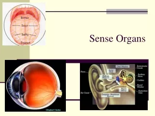
Nose: • The olfactory or smell action is quite simple. Once the smell of food has reached your nose, which is lined with hairs, it travels to an olfactory bulb, a set of sensory nerves. The nerve impulses travel through the olfactory tract, around, in a circular way, the thalamus, and finally to the smell sensory cortex of your brain, located between your eye and ear, where it is interpreted to be understood and memorized by the body.
Eye: • Seeing is one of the most pleasing senses of the nervous system. This cherished action primarily conducted by the lens, which magnifies a seen image, vitreous disc, which bends and rotates an image against he retina, which translates the image and light by a set of cells. The retina is at the back of the eye ball where rods and cones structures along with other cells and tissues covert the image into nerve impulses which are transmitted along the optic nerve to the brain where it is kept for memory.
Tongue: • A set of microscopic buds on the tongue divide everything you eat and drink into four kinds of taste: bitter, sour, salty, and sweet. These buds have taste pores, which convert the taste into a nerve impulse and sends the impulse to the brain by a sensory nerve fiber. Upon receiving the message, your brain classifies the different kinds of taste. This is how you can refer the taste of one kind of food to another.
Ear: • Most people only relate the ear drum or tympanic membrane as the only structure in the ear. However, this is not true. The cartilage over the cell ear is called the auricle. This area serves as a protective member to guard the inner ear where the famous ear drum is located. The typmanic membrane is divided into two layers. One that goes to the pharynx and the other is a mucus membrane. By putting pressure of both membranes is the only way the ear drum receives sound. Once the sound or sound wave has entered the drum, it goes to a large structure called the cochlea. In this snail like structure, the sound waves are divided into pitches. The vibration of the pitches in the cochlea are measured by the Corti. This organ transmits the vibrational information to a nerve, which sends it to the brain for interpretation and memory.
Function: • The main role of the respiratory system is to provide gas exchange between the blood and the environment. Primarily, oxygen is absorbed from the atmosphere into the body and carbon dioxide is expelled from the body.
Nose and Nasal Cavity: • As you inhale, small specks of dirt are trapped by many tiny hairs in your nose. This cleans the air. The hairs stop the dirt from going further in your body. The moist inside surface in your nose traps even smaller pieces of dirt. The nasal cavity, the air passage behind the nose, plays an important role in breathing. The nasal cavity is divided into a right and left passageway. The tissue that covers the wall of your nasal cavity contains many blood vessels. Heat from the blood in the vessels helps warm the air as you breath. Moisture is added to the air you breath by special cells in the walls of the nasal cavity. The air is warmed and moistened before it reaches your lungs.