Download
1 / 35
360 likes | 618 Views
Cancer. What is Cancer?. uncontrolled cell growth (as opposed to steady-state replacement of cells) usually accompanied by de-differentiation of cells cancerous mass = tumor or neoplasm Natural selection: cells which grow faster than others will take up more and more space.

E N D
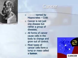
What is Cancer? • uncontrolled cell growth (as opposed to steady-state replacement of cells) • usually accompanied by de-differentiation of cells • cancerous mass = tumor or neoplasm • Natural selection: cells which grow faster than others will take up more and more space. • Our cells have multiple defenses against cells overgrowing their allotted locations. • Cancer occurs when those defenses have been removed. • starts with one transformed cell
Basic types of cancer • Sarcoma is derived from mesodermal tissue such as bone, muscle. • Carcinoma is derived from ectoderm or endoderm such as skin, nerves. • Leukemia is derived from white blood cells(WBC) from bone marrow. • Lymphoma is derived from WBCs from lymph glands.
Genetic Phenomenon • Cancer involves changes in DNA sequence i.e. it is genetic • Cancer is not epigenetic i.e. changes in patterns of gene expression without DNA changes. • If cancer were epigenetic, it might be easier to reverse. • Epigenetic changes, such as DNA methylation and histone modification, do occur in cancer, but they are rarely or never the underlying cause.
Why we think cancer is genetic: 1. Most mutagens are carcinogens: • Many carcinogens are activated in the liver: need to treat with liver extract before doing Ames test. • Ames test: his- Salmonella are reverted to his+ by mutagens. They will then grow on medium lacking histidine.
Why we think this (cont) 2. Tumors are clonal: • X-inactivation. • 2 alleles of Glucose 6-phosphate dehydrogenase, located on X. • Only 1 is expressed in any given tumor.
Why we think this (cont) 3. Cancer runs in families: • Survey of 4000 visitors to a general clinic, • 50% had little or no cancer in their families, • 7% had 3 or more close relatives with it. • Of the 7% of clinic visitors with 3 or more close relatives with cancer: • Pedigrees show that there are many types of cancer in these families • Implying a general propensity to cancer rather than a specific type
Types of Cancer in Families • Li-Fraumeni syndrome is a rare hereditary disorder that increases greatly the susceptibility to cancer. The syndrome is a mutation in the tumor suppressor gene, which normally helps control cell growth. • Xeroderma pigmentosum--lack of DNA repair in skin leads to skin cancers, esp. with sunlight exposure. • Also several others. --specific chromosome breakage syndromes e.g. ataxia-telangiectasia. Many breaks, tendency to breast cancer.
Cancer is a progressive disease • Needs 5-6 mutations for full-blown cancer. • Involves natural selection--in a slow-growing tumor, a faster growing mutant will take over.
Stages of Cancer • Initiation: A mutation that transforms the cell, leaving it capable of unrestrained growth. • Promotion: No growth unless cell enters S phase (many cells are arrested and need a promoter, a mitogen, to get them started) • Progression: • Angiogenesis--invasion of tumor by blood vessels • Invasiveness--ability to penetrate basal membranes. Tumors that can't do this are benign, those that can are malignant • Metastasis--ability to go through the blood and colonize other tissues
Cervical Cancer example • Cervix is a multi-layered tissue with a basal membrane. • Normally, all cell division occurs only in layer next to basal membrane. • Dysplasia - patches of cells multiply above the basal membrane. • Seen in Pap smear. • Often remains harmless and sometime reverts to normal. • With carcinoma in situ, (badly named--it is not full-blown cancer), all layers in an area are de-differentiated and dividing. • Easy to cure at this point with surgery. • Malignant carcinoma - crosses basal membrane and invades underlying connective tissue.
Tumor Inducing Viruses • Key to our current understanding of cancer. • DNA viruses--complicated systems: • Epstein-Barr virus --Burkitt's lymphoma • hepatitis B--Liver cancer • papilloma virus (genital warts)--cervical cancer • HTLV I and II --leukemia
DNA viruses • DNA viruses: normally replicate in the cell as plasmids, but occasionally integrate into the genome, where viral promoters can by chance activate oncogenes. • Very complicated genomes
RNA viruses • RNA viruses: retroviruses. • First one Rous sarcoma virus • Discovered by Peyton Rous in 1917 • Chickens could get cancer from a filterable extract of tumors. • structure: LTR--gag--pol--env--LTR • LTR--long terminal repeat. For integration and transcription (promoter) • gag—inner core RNA-binding proteins • pol--reverse transcriptase adn other enzymes • env--envelope glycoproteins (virus has membrane) • expression--interesting splicing and cleavage of RNA and polyproteins
Oncogenes • Two very similar retroviruses: RSV and ALV • Rous Sarcoma Virus is 10 kb and acutely transforming (tumors in weeks). • Avian Leukosis Virus is 8.5 kb and weakly transforming: latent period of months or more, and often has no effect. • temperature sensitive mutants of RSV: replicate at all temperatures, but transform at 35o but not 41o. That is, a genetic separation of transformation from replication--different genes involved. Temp sensitive mutants due to mutant protein that is unstable at high temps. • Transforming gene at 3' end of RSV. Called src.
Transforming Retroviruses • Most transforming retroviruses are replication-defective. • They only replicate in mixed infection with normal (non-oncogenic) retrovirus. e.g. Abelson MuLV has gag deleted at 3' end fused to abl oncogene, generating a fusion protein, and nothing else in genome. • source of viral oncogenes • Varmus + Bishop 1976 used a src probe against chicken genomic DNA in a Southern blot • got a band i.e. src is a normal cellular gene as well as a viral gene. • True of all viral oncogenes: • v-onc = viral version. No introns • c-onc = cellular version has introns • v-oncs are also often mutated, partially deleted, or fused to other sequences as compared to c-oncs. • presumably got into viruses by viral genome integrating next to c-onc and a mistake occurring allowing c-onc to be transcribed along with viral genome. • A certain amount of mutation at this point would give a v-onc surrounded by LTRs. • In the cell, oncogenes do not normally cause cancer, only their mutated versions do. The normal, non-carcingeneic versions are often called “proto-oncogenes”.
Gene Transfer Experiments • Gene Transfer experiments are another approach to identifying oncogenes • Showed that oncogenes in human tumors are the same as oncogenes identified from retroviruses. • Normal fibroblasts will multiply in Petri dishes, but they have 2 specific properties of interest: • contact inhibition: they stop growing when they touch, leading to a monolayer. • finite number (50-60) of cell divisions before death
Partially Transformed Cells • When transformed, cells lose contact inhibition (they pile up) and become immortal. • NIH 3T3 mouse cells are partially transformed: immortal but still contact inhibited. That is, they grow in a monolayer • However, mutagens, etc. will create foci (plural of focus) of piled up cells starting with a single transformed cell. • Basis for oncogene assay.
The Experiment • --extract DNA from bladder carcinoma EJ. • Use to transfect 3T3 cells (CaPO4 precipitation induces pinocytosis). See foci of transformed cells, meaning that some of the human DNA can transform. • Non-tumor DNA does not transform. • now, extract DNA from foci: contains mostly mouse DNA with just a bit of human containing the oncogene. • Re-transfect fresh 3T3 cells, get new foci. This removes more of the extraneous human DNA. • Clone DNA from the foci, screen with Alu sequence • Alu sequences are very common throughout the human genome and thus are linked to most human genes. Not found in mouse DNA at all. • Then sequence the resulting clones. • Turned out to by H-ras, already known from Harvey rat sacroma virus • had one mutation: Gly converted to Val • other human tumors processed this way yield other oncogenes. • but not all work.
Activating Oncogenes • Normally, cellular oncogenes are proto-oncogenes: they have a regular cellular function and aren’t involved with cancer. • Two basic ways of converting proto-oncogenes into oncogenes: • mutate the protein • make lots of the normal protein • There are a variety of ways to accomplish these events.
Types of Mutation that Create Oncogenes • Base changes and other simple alterations in protein structure. e.g. H-ras described above as the cause of bladder carcinoma in the transfection experiments. • Creation of a fusion protein: • Starts out a normal (often highly expressed) cellular protein • Ends up with oncogene sequences, creating a N-terminal deleted oncogene with other sequences added. • This can have abnormal function. • Created by translocation (e.g. Philadelphia chromosome (short 22--actually a t(9;22))that joins c-abl with bcr (breakpoint cluster region) gene. • Creates an unusually active Abl protein that causes chronic myeloid leukemia.
Types of Mutation that Create Oncogenes (cont.) Types of mutation to create active oncogenes from proto-oncogenes: • Replacing normal weak promoter of proto-oncogene with strong promoter, leading to over-expression. • Insertional mutagenesis: the LTR is often a strong promoter for sequences 3' to it. • The cause of oncogenesis in weakly oncogenic viruses like ALV. • ALV often inserts in c-myc gene just before protein-coding sequences, giving lots of normal Myc protein. • Gene amplification. • Tumors often become insensitive to chemotherapeutic agents due to gene amplification: many duplications of target gene. • Classic case is methotrexate resistance due to amplification of dihydrofolate reductase gene. • Gives homogeneously staining regions of chromosome due to many tandem copies and/or double minute chromosomes, containing copies of the gene but no centromere. • Many tumors have this: • myc in lung cancer • erb -B2 in breast and ovarian tumors • Get hundreds of copies of the gene • Comparative genome hybridization: label DNA from normal cells and from tumor cells with red and green fluorescent tags. Hybridize to normal chromosomes and scan to see red/green ratio. Deviations show regions of amplification or loss in the tumor line.
Tumor Suppressor genes • A distinction: • Oncogenes act in a dominant fashion: one mutant copy plus one normal copy gives a tumor. • Tumor suppressor genes are recessive: one mutant and one normal is still wild type--need both copies mutant to give a tumor. • Wild-type oncogenes (proto-oncogenes) promote cell proliferation; mutant versions enhance this property. • On the other hand, tumor suppressors regulate and inhibit cell proliferation; mutant versions remove controls on proliferation. • Tumor suppressor genes mostly found by cloning familial cancer genes and chromosome regions commonly deleted in tumor cells.
Retinoblastoma • Retinoblasts are retinal precursor cells that are only found in young children. • Inherited and spontaneous forms of retinoblastoma. • Inherited form is autosomal dominant and usually both eyes are affected. • Spontaneous is almost always one eye only. • Theory of Knudson (1971): both copies of the Rb gene need to be inactivated by mutation to give a tumor. • Inherited form: • People start with one copy knocked out: RB+ RB- • They thus need only a single mutation to get a tumor. • Mutation rate is around 1 in 106; • 8-10 million cells per retina means that at least one cell in both retinas usually gets hit and forms a tumor. • Spontaneous: • Need 2 hits, • Each with a 10-6 rate, or 1 in 1012--quite rare.
How to Lose a Gene • Many events leading from an RB heterozygote to cancer are not classical mutations, but rather "loss of heterozygoisty" (LOH), where one copy of the gene is deleted or inactivated, or converted to the same allele as the other copy. • Various causes: spontaneous mutation or deletion, non-disjunction, mitotic recombination. Inactivation by methylation of the non-mutant copy also occurs. • Sometimes can see deletion or loss of marker loci in region of the RB gene (13q14) in tumor cells.
Why is RB a dominant trait? • Semantics. • At the whole body level, it is dominant because you need to inherit it from only 1 parent. • At the cellular level, it is recessive because both copies need to be knocked out to get the transformed cell. • A Rb homozygote would probably be dead.
Other Cases • Wilm's tumor. • Part of WAGR syndrome (Wilm's, aniridia, genital abnormalities, retardation) found in heterozygous deletion of 11p. • Homozygous deletion is dead as are virtually all homozygous deletions of more than 1 or 2 genes. • Apparently 4 separate genes involved in syndrome. • WT is childhood kidney tumor same deal as RB • spontaneous is one side only • inherited is usually bilateral • Li-Fraumeni syndrome: loss of p53, a 53 kdal protein that is the “central guardian of the genome”, preventing replication or killing cells that have DNA damage. • Loss of 17p region where p53 is seen in many tumors: colon, lung breast etc. • Ataxia telangiectasia. The disease has many symptoms: neurodegeneration, immunodeficiency, premature aging, radiation sensitivity • The ATM protein is part of the pathway that signals DNA damage • cloned by using cosmids with normal DNA to rescue an AT cell line from its radiation-sensitive phenotype
What are oncogenes and tumor suppresors? • A lot of fundamental cell processes have been investigated as part of understanding oncogenes. • Several basic types: • growth factors • growth factor receptors at the cell surface • signal transduction proteins • transcription factors • cell cycle regulatory proteins • DNA damage detection and repair proteins
Growth Factors / Analogs • Some oncogenes are growth factors or analogs. e.g. sis is very similar to PDGF (platelet-derived growth factor) • If turned on in a cell carrying PDGF receptors, it will self-stimulate to unrestrained growth. • Normally PDGF is made in one cell and the receptors are on other cells • Opposite side of this: some oncogenes are growth factor receptor analogs: • e.g. erbB is a mutated version of epidermal growth factor receptor (EGF). • Normally EGF receptor is a transmembrane protein that binds EGF on outside and a kinase on the inside that is activated only when EGF is bound outside membrane. • erbB oncogene deletes most of the outside part, leading to a kinase that is always on. • Just like continuous application of EGF--continuous growth and division.
Intracellular Transducers and Transcription Factors • Some oncogenes are intracellular transducers • Lots of targets: lots of pathways and interactions • e.g. src, ras, abl • Some oncogenes are transcription factors: • cause sets of genes to be activated or inactivated inappropriately. • e.g. fos/jun combine to form factor AP1, which binds to a specific DNA sequence for transcription.
Cell Cycle Control • Complex and not fully understood yet. Many overlapping control systems • In general a cell can: stay in interphase, divide, or undergo programmed cell death (apoptosis). • Checkpoints: the cell cannot proceed past them until certain conditions are met. • G1 -> S • G2 -> mitosis • metaphase spindle attachment • G1-S checkpoint. The main control point for cells with damaged DNA • the main control protein is pRb (product of RB1, the retinoblastoma gene). • It has 2 states: phosphorylated (inactive) and dephosphorylated (active). • pRb acts on the transcription factor E2F1. If E2F1 is active, the cell can move from G1 phase to S phase, the first step in getting the cell to divide. • The role of pRb is to inhibit E2F1: when pRb is active (dephosphorylated), E2F1 is inactivated, so the cell stays in G1. • If pRb is phosphorylated (by a cascade of kinases), it becomes inactive, and E2F1 becomes active; the cell then enters S phase. • Phosphorylation of pRb is done by cyclins (proteins whose concentration varies with the cell’s position in the cell cycle) and CDKs (cyclin-dependent kinases). • G2-M checkpoint. Cells must have completed DNA repair to pass this point • Mitosis is initiated by the MPF (maturation promoting factor) protein complex, composed of cyclins and CDKs which have built up over the course of the cell cycle.
Genome Integrity • Mutation is a constant problem. Many mechanisms prevent cells with seriously mutated DNA from dividing. • Malignant cells usually undergo chromosomal rearrangements, leading to new fused genes and loss of heterozygosity. • Spindle checkpoint. During mitosis, cells can only proceed into anaphase when all of the chromosomes are properly attached to the spindle. A protein complex on the kinetochore is displaced when the kinetochore is attached to the spindle. • Telomerase. This enzyme prevents the loss of DNA at the ends of chromosomes, an inevitable consequence of replication. It is inactive in most cells, which results in them dying after 60 or so cell divisions. However, it is re-activated in 85% of successful tumor cells, resulting in cellular immortality. This is one of the most common markers of cancer.
DNA Damage Detection • Several DNA repair systems are tumor suppressor genes. • BRCA1 and BRCA2: genes implicated in breast cancer. Also ATM, the ataxia telangiectasia protein. Part of a multi-protein BASC complex that scans the DNA for damage • Xeroderma pigmentosum: DNA damage caused by sunlight isn't repaired, leading to skin tumors. • Several forms, involving nucleotide excision DNA repair enzymes. • Hereditary non-polyposis colon cancer (doesn't form polyps). • Autosomal dominant • When the genomes of HNPCC patients were scanned for LOH, many micro-satellite loci had changes in repeat number, all over the genome. • Related to E. coli mutator system MutHLS, • Mutations in these genes increase mutation rate in E. coli up to 1000 x. • Mismatch repair system: removes mismatched DNA bases on newly synthesized strand and re-synthesizes that stretch of DNA. • Human analogue of MutS gene mapped to region of HNPCC gene, and turned out to be mutant in HNPCC patients. • One human homologue of MutL is also responsible for much of HNPCC
p53 and Apoptosis • p53 is the central regulator of genome integrity, “the guardian of the genome”. • p53 is a transcription factor, the protein product of the TP53 gene. • In normal cells, the p53 level is low because p53 is rapidly degraded by being ubiquitinated and processed by the proteosomes. • DNA damage and other stresses cause p53 to become phosphorylated, which stabilizes it. • This leads to a buildup of p53, which causes transcription of genes that inhibit the cell cycle and also initiate apoptosis. • Mutations in TP53 are very common, especially in late stage tumors. • It is the gene mutated in Li-Fraumeni syndrome, which causes a general increase in all kinds of tumors. • Also mutated in cigarette smoke-induced tumors, sunlight-induced tumors, aflatoxin-induced tumors, cervical cancer (papilloma virus-induced). • Apoptosis is programmed cell death: a signal (internal or external) activates a mechanism that kills the cell. • Very important is embryonic development • Cells with DNA damage undergo apoptosis • Cancer often develops due to a failure of apoptosis. • The apoptosis process is carried out by a set of proteases called caspases.
Cancer as Multi-step Process Progression of colorectal cancer (familial adenomatous cancer) • Adenomas are polyps seen in the colon. They start as abnormal crypt cells, progress to benign polyps, and finally become cancerous. • The following shows one way an FAP case may develop: • Normal colon epithelium has mutation in APC tumor suppressor gene causing rapidly proliferating epithelium. • Activation by mutation of KRAS (ras-K) leads to polyp formation. • Loss of heterozygosity tumor suppressor gene in 18q (exact gene not clear) leads to late stage polyp. • Mutation in p53 leads to carcinoma.