Download
1 / 37
380 likes | 536 Views
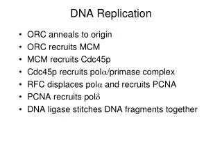
DNA Replication: Clamp Loaders and Other AAA+ Machines Within the Replisome Scott Morrical. Clamp Loader Review: g -complex of E. coli DNA Polymerase III. Loading of b 2 Clamp by g -complex ( g 3 dd ’), an AAA+ Machine. Structural Organization of Pol III Holoenzyme.

E N D
DNA Replication: Clamp Loaders and Other AAA+ Machines Within the Replisome Scott Morrical
Clamp Loader Review: g-complex of E. coli DNA Polymerase III
Loading of b2 Clamp by g-complex (g3dd’), an AAA+ Machine
Structural Organization of Pol III Holoenzyme
AAA+ Proteins: ATPases associated with a variety of cellular activities
AAA+ ATPases (E. coli g protein shown) • Typically 2 domains: • Domain I-- boxes II - VII • Domain II-- boxes VII’ - Sensor 2 • Some of the AAA+ sequences are found in many other NTPases, with those sequences beyond Sensor-1 and to the end of Sensor-2 motif defining the AAA+ family. • Box VII motif (dark blue) contains a highly conserved Arg residue (R169) that may function analogously to “arginine finger” of GAP proteins. • Sensor-2 motif (purple), unique to AAA+ family, forms an a-helix across top of ATP-binding site analogous to “lid” segment of adenylate cyclase.
AAA+ ATPases (E. coli g protein shown) • Sensor-1 motif (medium blue) contains a conserved Thr residue (T155) that may interact with g-phosphate of ATP, analogous to the Switch II helix in G proteins. • Also a conserved Arg residue (R215) in Sensor-2 motif (purple) may detect the presence of ATP g-phosphate. • Walker-A (orange) and Walker-B (cyan) motifs are found in many NTPases. These are mutated in AAA+ proteins to prevent interactions with ATP (e.g. d and d’ proteins of g-complex and Orc4 or ORC). • AAA+ core is found in many other NTPases and is similar to bacterial RecA recombinase-- an a,b-fold that includes the sequences from Walker-A (P-loop) to the Sensor-1 motifs.
AAA+ Proteins Involved in DNA Replication Bacteria g3dd’ clamp loading DnaA initiator DnaC helicase loader Eukaryotes RFC clamp loading Orc1, Orc4, Orc5 initiators Cdc6 initiation, helicase loader? Mcm2-7 replicative helicase?
Structural Biology of E. coli g-complex Jeruzalmi, D., O’Donnell, M., and Kuriyan, J. (2001) Crystal structure of the processivity clamp loader gamma (g) complex of E. coli DNA polymerase III. Cell 106, 429-441. Kazmirski, S.L., Podobnik, M., Weitze, T.F., O’Donnell, M., and Kuriyan, J. (2004) Structural analysis of the inactive state of the Escherichia coli DNA polymerase clamp-loader complex. Proc. Natl. Acad. Sci. USA 101, 16750-16755. Jeruzalmi, D., Yurieva, O., Zhao, Y., Young, M., Stewart, J., Hingorani, M., O’Donnell, M., and Kuriyan, J. (2001) Mechanism of processivity clamp opening by the delta subunit wrench of the clamp loader complex of E. coli DNA polymerase III. Cell 106, 417-428.
Structure of the g-complex (nucleotide-free form) C-terminal collar (domain III) N-terminal AAA+ (domains I & II) front view of g subunits only top view bottom view
Gamma-complex = heteromeric pentamer • All 5 AAA+ subunits are in different conformations with respect to each other. • Domains I & II comprise AAA+ core of each subunit. (The upper panel shows each subunit with the AAA+ core in equivalent orientation.) • C-terminal Domain III is unique to clamp loaders among AAA+ proteins of known structure. Forms helical scaffold or “collar”. Domains: III II I
Structures of g Subunits • Here they are shown as if C-terminal domains are overlaid, emphasizing their different conformations, which involves different degrees of rotation between Domains I, II, & III. • g subunits are the ATPase motor subunits of the complex. Although complex was crystallized without ATP, it has been modeled in based on comparisons with other AAA+ protein-ATP complexes of known structure. • d’ is a conformationally stable “stator”, whereas d is a conformationally flexible “wrench” that binds independently to b subunit. d and d’ do not bind or hydrolyze ATP. • Panel B compares the nucleotide binding regions of g3 and d’ subunits. The side chain of Arg215 from the sensor region of g3 is shown. In d’the nucleotide binding site is blocked by an N-terminal extension, of which Met1 is one of the residues forming a conserved hydrophobic patch on the surface (replaced by ion pair in g3).
The C-terminal Domains of g, d, and d’ Form a Helical Scaffold • The C-terminal domains of d’ and the 3 g subunits interact with each other in a similar manner, in the order d’- g1- g2- g3. • The C-terminal domain of d is bound between the g3 and d’, closing the circle, but it is displaced outward from the circle and has different interfaces with both g3 and d’. • This asymmetry is propagated into to AAA+ domains of d, which includes the b interaction element.
Nucleotide Binding by g-complex • There are 3 different potential nucleotide binding sites in the complex, each located near an interface. • In each pair of subunits, the Sensor 1 region of the first subunit is positioned near the ATP binding site of the next one. • Although d’ does not bind nucleotides, it has the conserved Sensor 1 SRC motif, and this is positioned near the binding site of g1. This arrangement is followed sequentially by g2 and g3. • The sensor 1 region of g3 does not abut a nucleotide binding site since one is lacking in d. Instead, g3 SRC motif is involved in binding Domains I & II of d tightly to Domain I of g3.
Nucleotide Binding by g-complex (cont’d) • g1 nucleotide binding site (at d’- g1 interface) is open. • g2 nucleotide binding site (at g1- g2 interface is closed and Sensor 1 motif of g1 is deeply buried. • g3 nucleotide binding site (at g2- g3 interface) is open and causes the d wrench to be swung out. • The open-closed-open arrangement of g subunit nucleotide binding sites is reflected in crystal structures of the complex obtained in the presence of ATPgS and ADP. • Both nucleotides co-crystallize with g-complex with a 2:1 stoichiometry. • One nucleotide binds to the g1 nucleotide binding site (at d’- g1 interface), and one to the g3 nucleotide binding site (at g2- g3 interface). • This open, unsaturated structure is thought to represent the stable, inactive state of the complex, in which the ATPase domains are prevented from fully engaging the clamp.
X-ray Structure of bd Complex View along edge of b ring, centered on Domain 3 of b b interaction element of d (in yellow) consists of helix a4 in the N-terminal domain (Domain I) plus the loop following it. View showing intermolecular interface involving Domain 3 of b
Molecular dynamics simulations of ns timescale dynamics of b conformational change suggests that b2 dimer may be “spring-loaded”, with potential energy stored in strong inter-dimer interface. d either induces a conformational change in b that trips the spring, or captures a spontaneously ring-opened form.
Structural Biology of Yeast RFC-PCNA Complex Bowman, G.D., O’Donnell, M., and Kuriyan, J. (2004) Structural analysis of a eukaryotic sliding DNA clamp-clamp loader complex. Nature 429, 724-730.
Stable Interaction of Yeast RFC Complex with a Closed PCNA Ring • Closed, active conformation of clamp loader contains 5 bound nucleotides (e.g. ATPgS) even though RFC-E (aka RFC5 = d’ = stator) lacks several conserved ATPase residues.
RFC subunits in complex form right-handed spiral array, the formation of which is dependent on ATP (gS) • RFC-A (aka RFC1 = d = wrench) contributes a unique C-terminal Domain IV, which is situated between the ATPase domains of RFC-A and RFC-E, and provides a physical link between the two ends of the RFC spiral. • PCNA has 3 conserved hydrophobic grooves for potential interaction with RFC, 2 of which are enganged by RFC-A and RFC-C.
A Model for Primed DNA Interacting with RFC-PCNA Complex • Spiral RFC complex, like a screw-cap, threads onto the last turn of the duplex, further extension of which is blocked by the C-terminal collar. This imposes directional specificity on complex-DNA interactions. • There appears to be a path for the 5’ ssDNA end of the template strand to snake out of the complex. • DNA polymerases interact with the same face of the sliding clamp as do the clamp loaders, so the clamp is now positioned correctly for DNA synthesis upon departure of RFC.
Conserved residues in Domain I of clamp loader subunits at the proposed DNA-interacting surface. Loops preceding the central (a5) and SRC (a6) helices are equivalent to the L1 and l2 loops of RecA, which have been implicated in DNA binding through extensive mutagenesis of RecA and DnaB-type helicases.
Inactive and Active Clamp Loader Complexes • Nucleotide-free, inactive state of E. coli clamp loader appears to form a proto-spiral • Nucleotide-bound, active state of yeast clamp loader forms a tight spiral that complements the helical repeat of primer-template
Other AAA+ Machines in the Replisome
AAA+ Tau Links Leading, Lagging Strand Polymerases, & Clamp Loader in Pol III Holoenzyme
Initiation of E. coli DNA Replication at oriC-- roles of DnaA Initiator Protein and DnaC Helicase Loader DnaA-- initiator protein oriC-- replicator sequence
Erzberger, J.P., Pirruccello, M.M., and Berger, J.M. (2002) The structure of bacterial DnaA: implications for general mechanisms underlying DNA replication initiation. EMBO J. 21, 4763-4773.
Alignment of DnaA sequences from E. coli and from thermophilic bacterium Aquifex aeolicus. • Domain I = N-terminal region involved in DnaB helicase loading at oriC. • Domain II = extended linker segment. • Domain III = AAA+ ATP binding cassette. • Domain IV = DNA binding domain recog- nizing 9 bp repeat sequences in oriC.
Crystal structure of DnaA-ADP complex from thermophilic bacterium Aquifex aeolicus • Truncated protein containing only Domains III & IV. • Biochemical data indicates that DnaA oligomerization and DNA binding/ remodeling functions reside in Domains III & IV. • Note Domains IIIa and IIIb represent Domains I and II, respectively, of classic AAA+ fold.
Known mutations of E. coli DnaA mapped onto A. aeolicus structure • DNA binding domain resembles classic HTH motif such as found in Trp repressor • Highly conserved residues in the basic loop and DnaA signature sequences are indicated by spheres. • Residues determineed by mutagenesis to be critical for DNA binding in E. coli that map to HTH and basic loop motifs are highlighted in red.
Model for DnaA Binding to oriC • oriC is recognized by DnaA (red, green, yellow) and by architectural factors such as IHF and HU (purple). • Concomitant with oriC binding, the AAA+ domains oligomerize, stabilizing the nucleoprotein complex through inter-monomer contacts around the ATP binding site. Additional stability may be provided by domain I self-oligomerization (light blue). Self-assembly of DnaA molecules eventually leads to formation of the complete nucleoprotein complex. • The DnaA oligomer could conceivably accommodate either a closed ring (left) or a helical filament (right) arrangement of monomers. • DUE opening may occur spontaneously through local strain induced by assembly of the nucleoprotein complex in the presence of ATP.
Shameless Speculation About Helicase Loading Mechanisms at Eukaryotic Origins