Download
1 / 23
270 likes | 1.4k Views
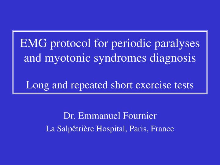
EMG protocol for periodic paralyses and myotonic syndromes diagnosis Long and repeated short exercise tests. Dr. Emmanuel Fournier La Salpêtrière Hospital, Paris, France. Overview of the protocol.

E N D
EMG protocol for periodic paralyses and myotonic syndromes diagnosisLong and repeated short exercise tests Dr. Emmanuel Fournier La Salpêtrière Hospital, Paris, France
Overview of the protocol • The EMG protocol includes several tests performed successively or simultaneously in order to reduce the total examination duration : • Long (5 mn) exercise test on right hand, with post-exercise rest • Repeated short (10s, three times) exercise test performed two times, on left hand and on right foot • Needle electromyography of five muscles • References for the precise description of technical conditions, protocol and main results : • Fournier, E.M.Inter, Cachan, 1998, pp 253-262 • Fournier et al. Annals of Neurology, 2004, 56: 650-661 • Fournier, Rev Neurol (Paris), 2005, in press
Preparation • Three sets of surface recording electrodes are installed • Best stimulation and recording sites are located • Baseline compound muscle action potentials (CMAP) are recorded • Recording electrodes are left in the optimal position throughout the examination • The purposes are threefold : • Electrodes will serve to different exercise tests • A rapid nerve conduction study checks the absence of nerve alterations. If necessary, neuromuscular transmission is tested by 3 Hz repetitive nerve stimulation. • Baseline recordings would serve as reference values if an attack of paralysis occurs during the examination
Preparation (I, II, III) • Recording of the extensor digitorum brevis (EDB) muscle • Stimulation of the anterior tibial nerve • Electrodes will be used for one of the repeated short exercise tests • Recording of the left abductor digiti minimi (ADM) • Stimulation of the ulnar nerve • Electrodes will be used for one of the repeated short exercise tests • Recording of the right abductor digiti minimi (ADM) • Stimulation of the ulnar nerve • Electrodes will be used for the long exercise test
Preparation (IV) • A bandage prevents articulation displacements and changes in muscle volume during the exercise tests • Skin temperature is measured and must be maintained between 32 and 34°C throughout the EMG session • The purpose is to prevent any decrease in CMAP amplitude and area which could be caused by muscle warming during the examination
Long exercise test on right hand (I) • CMAP is monitored before exercise (top trace) • The subject is asked to spread the little finger as strong as possible against resistance during 5 minutes • Brief resting periods (2-3 seconds) are respected every 30-45 seconds to prevent ischemia • After completion of the exercise, the subject is instructed to completely relax while CMAP are recorded • 2 seconds immediately after cessation of exercise (second trace) • and then 30 seconds after (third trace)
Long exercise test (I)Normal changes immediately after exercise • Immediately after exercise cessation, normal results are : • Slightly decreased amplitude (-6 %) of the CMAP • Increased duration (+38 %) disappearing in 30 seconds • If the amplitude decline is more marked, it must be carefully checked that nerve stimulation is still supramaximal
Long exercise test (I)Immediate changes after exercise cessationin muscle channelopathies • Patients with Paramyotonia Congenita • Decreased CMAP amplitude • Presence of Post-exercise myotonic potentials (PEMP) • Patients with Periodic Paralysis • No change of the CMAP in most cases • Early decreased amplitude if an attack of paralysis has been triggered by the exercise achievement • Increased amplitude in patients with Hyperkalemic Periodic Paralysis, especially when pre-exercise CMAP amplitude was small
Long exercise test (II)Changes 1-5 minutes after exercise • CMAP is recorded every minute during the first 5 minutes after exercise cessation (traces 4-8 on the screen) • Normal results during this rest period are no change of the CMAP as compared with pre-exercise values : • Otherwise, it must be carefully checked that nerve stimulation is still supramaximal
Long exercise test (II)Changes 1-5 minutes after exercisein muscle channelopathies • Patients with Paramyotonia Congenita • Disappearance of PEMP, persistence of decreased CMAP amplitude • Patients with Periodic Paralysis • No change or progressive decline of CMAP amplitude • Persistence of the increased amplitude if previously present, in Hyperkalemic Periodic Paralysis
Continuation of the long exercise test • CMAP of the right ADM is recorded every 5 minutes during the 40 minutes rest after exercise cessation. • Recording electrodes and bandage are left on the right hand • During the 5 minutes interval between two recordings, different others tests are performed : • Repeated short exercise test on the left hand • Repeated short exercise test on the right foot • Search for myotonic discharges by needle-EMG
Repeated short exercise teston left hand (I) • A second bandage is installed on the left hand, and CMAP of the ADM is monitored before exercise (top trace) • The subject is asked to spread the little finger as strong as possible against resistance during 10 seconds • After completion of the exercise, the subject is instructed to completely relax while CMAP are recorded at rest : • 2 seconds immediately after cessation of exercise (second trace) • and then every 7-8 seconds for 50 seconds (traces 3-8 on the screen) • This sequence (10 s contraction - 50 s rest) is repeated three times
Repeated short exercise test (II)Normal CMAP changes after exercise • Immediately after exercise cessation, normal results are : • Slightly increased amplitude (+5 %) of the CMAP (second trace) • Return to pre-exercise values in 10 seconds (third and next traces) • If an amplitude decline appeared, it must be carefully checked that nerve stimulation is still supramaximal
Repeated short exercise test (II)CMAP changes between the exercisesin muscle channelopathies • Patients with myotonic syndromes • Presence of Post-exercise myotonic potentials (PEMP) immediately after the first short exercise, especially in Paramyotonia Congenita • Decrease in CMAP amplitude, worsening or disappearing with repeating exercise in Paramyotonia Congenita and Myotonia Congenita respectively • Patients with Periodic Paralysis • No change of the CMAP in most cases • Increased amplitude in patients with Hyperkalemic Periodic Paralysis, worsening with repeating exercise, especially when pre-exercise CMAP amplitude was small
Long exercise test (III) • Back to the right hand for recording CMAP of the ADM at 10 minutes rest after exercise cessation. • If an amplitude decline appeared, it must be carefully checked that nerve stimulation is still supramaximal
Repeated short exercise teston right foot • CMAP of the EDB is monitored before exercise (top trace) • The subject is asked to raise the toes as strong as possible against resistance during 10 seconds • After completion of the exercise, the subject is instructed to completely relax while CMAP are recorded at rest : • 2 seconds immediately after cessation of exercise (second trace) • and then every 7-8 seconds for 50 seconds (traces 3-8 on the screen) • This sequence (10 s contraction - 50 s rest) is repeated three times
Long exercise test (IV) • Back to the right hand for recording CMAP of the ADM at 15 minutes rest after exercise cessation. • If an amplitude decline appeared, it must be carefully checked that nerve stimulation is still supramaximal
Needle Electromyography • EMG recording of 5 muscles at rest and during voluntary contraction : • Deltoid • Extensor digitorum brevis • First interosseus dorsalis • Tibialis anterior • Vastus medialis • Searching for myotonic discharges : • Abundant in myotonic syndromes • Rare and inconstant in Hyperkalemic Periodic Paralysis • Absent in Hypokalemic Periodic Paralysis
End of the long exercise test (V) • Back to the right hand for recording CMAP of the ADM at 20, 25, 30, 35, 40 minutes rest after exercise cessation. • Changes in CMAP amplitude between -20 and +10 % of the pre-exercise value can be considered normal. • If an amplitude decline appeared, it must be carefully checked that nerve stimulation is still supramaximal
End of the long exercise test (V)Changes 20-40 minutes after exercisein muscle channelopathies • Patients with Paramyotonia Congenita • Progressive disappearance of the immediate post-exercise decline in CMAP amplitude • Patients with Periodic Paralysis • Progressive installation of a marked decline of CMAP amplitude of the right ADM • In some cases, a paralysis of the four limbs occurs during the post-exercise rest, with marked CMAP amplitude declines not only of the right ADM, but also of the left ADM and of the EDB.
Long exercise test in periodic paralyses CMAP changes following long exercise in an unaffected patient (A), a patient with hyperkalemic periodic paralysis (hyperKPP) and T704M sodium channel mutation (B), a patient with hypokalemic periodic paralysis (hypoKPP) and R528H calcium channel mutation (C), a patient with Andersen-Tawil syndrom (ATS) and T309I potassium channel mutation (D). Pre-exercise (top trace) and post-exercise recordings (below) at different times following the trial (Ex) as indicated left to the traces. Scale between 2 dots: 5 ms, 5 mV. (Adapted from Fournier et al. Ann Neurol 2004, Fournier Rev Neurol 2005,Bendahhou et al. J Physiol Lond 2005)
Conclusion • Total duration of the protocol realization : 50 minutes • Preparation : 5 mn; Long exercise : 5 mn; Rest after long exercise with repeated short exercises and needle EMG : 40 mn • By comparing EMG findings and responses to long and repeated short exercise tests, five main patterns of muscle abnormalities could be defined (Type I-V), each of them corresponding to a defined group of ion channel mutations. • References for the results : • Fournier et al. Annals of Neurology, 2004, 56: 650-661 • Fournier, Rev Neurol (Paris), 2005, in press
Ackowledgments • Thanks to Ghislaine Therme for technical assistance and Nicolas Danziger for filming and comments. • This video tape was ordered and supported by Periodic Paralysis Association. • The work on muscle channelopathies was supported by Résocanaux and Association Française contre les Myopathies (AFM).