Download
1 / 28
510 likes | 1.12k Views
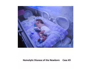
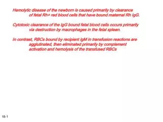
Hemolytic disease of newborn (HDN) " Erythroblastosis fetalis ". HDN result from transplacental passages of maternal antibodies (IgG) against RBC antigens of the infants, resulting in increase rate of RBCs destructions.

E N D
Hemolytic disease of newborn (HDN)"Erythroblastosis fetalis"
HDN result from transplacental passages of maternal antibodies (IgG) against RBC antigens of the infants, resulting in increase rate of RBCs destructions. It's an important cause of anemia and jaundice in newborn infants, the disease is associated primarily with the D-antigen of Rh group of RBC and with ABO groups incompatibilities, rarely, its might associated with minor antigens of RBC like C or E or "kell" antigens.
HDN due to Rh incompatibility: Pathophysiology: Its occurs if the mother have Rh negative blood group and the father (then the infant) have Rh positive blood group.
When Rh positive blood is infused in to Rh negative women through an error, or when small amount (usually >1ml) of Rh positive fetal blood containing D-Ag inherited from Rh positive father enter the maternal blood during pregnancy, abortion, or at delivery, this will induce AB formation against D-Ag in the mother circulation which is IgG type which is capable of passing through placenta during next pregnancy causing fetal RBC hemolysis by Ag-AB reaction.
HDN due to Rh incompatibility is rarely occur during the first pregnancy because transfusion of Rh positive blood in to Rh negative mother tend to occur near the time of delivery, which is too late for the mother to become sensitized and transmit Abs to her fetus before delivery. The severity of Rh sensitization increases with successive pregnancies, so the 1st affected infant after sensitization may represent the end of the end of the mother childbearing.
In summary Rh +ve blood → Rh –ve mother circulation → Abs formation against D-Ag of Rh +ve blood in mother circulation → cross placenta to fetal blood during next pregnancy → causing hemolysis of fetal RBCs.
Clinical features: The severity of Rh incompatible disease depends on the severity of hemolysis of fetal blood and its might be: 1. Only laboratory data of mild hemolysis (15% of cases). 2. Jaundice (Icterus neonatarum): Its might be absent at birth because of placental clearance of lipid-soluble unconjucated bilirubin, but in sever cases; bilirubin pigments stain the amniotic fluid, cord, and placenta by yellow color.
3. Hydrops fetalis: Its characterized by sever anemia, massive hepatosplenomegally, signs of cardiac failure (cardiomegally, respiratory distress), generalized edema (anasarca), circulatory collapse, and hypoalbuminemia due to liver dysfunction. Hydrps fetalis occur when fetal Hb level is < 5gm/dl. 4. Hypoglycemia: Occurs due to hyperinsulinism and hypertrophy of pancreatic Islet cells.
Laboratory data: 1. CBC shows: A. Anemia, which might be mild or sever with Hb, levels as low as 3-4g/dl B. WBC count normal or elevated. C. Thrombocytopenia in sever cases due to decrease production or DIC. 2. Peripheral blood smear shows polychromasia and marked increase in nucleated RBCs. 3. Increase reticulocytes count (reticulocytosis).
4. Strongly positive direct Coombs test. 5. Increased bilirubin level in cord blood (3- 5mg/dl) and unconjucated bilibirun increased rapidly to high levels in the 1st 6hr of life. Conjucated (direct) bilirubin may elevate also.
Postnatal diagnosis of Rh-hemolytic disease: Immediately after birth of any infant to Rh –ve women, blood from umbilical cord or from infant peripheral vein should be examined for: 1. ABO blood group and Rh type. 2. Hematocrit and Hb levels. 3. Direct Coombs test reaction. 4. Serial measurement of serum bilirubin levels.
Treatment of hydrops fetalis: 1. Immediate resuscitation and supportive therapy. 2. Temperature stabilization. 3. Correction of acidosis with 1-2 mEq/kg Na. bicarbonate. 4. Small transfusions of packed RBCs to correct anemia (5-20ml/kg). 5. Volume expanders to correct hypotension (NaCL, Ringer lactate or plasma). 6. Assisted ventilation for respiratory failure. 7. Exchange transfusion for treatment of hyperbilirubinemia.
Prevention of Rh sensitization: Prevention of initial sensitization of Rh –ve mothers could be achieved by: 1. I.M. injection of 300 Mg of human anti-D globulin (1ml) with in 72 hrs of delivery, ectopic pregnancy, abdominal trauma in pregnancy, amniocentesis, chorionic villus biopsy, or abortion. This amount of Anti-D globulin is sufficient to eliminate about 10 ml of potentially antigenic fetal cells from maternal circulation.
2. Avoidance of obstetric procedures that increase the risk of fetal-to-maternal bleeding e.g. fetal versions, manual separation of placenta and so on.
HDN due to ABO incompatibility: Pathogenesis: Major blood groups incompatibility between mother and fetus usually result in milder disease than does Rh incompatibility. Maternal Abs may be formed against B cells if the mother is type A or against A cells if the mother is type B, but in most cases the mother is type O and the fetus is type A or B. The Abs against A and B factors occurs without prior immunizations (natural Abs), but its IgM type which does not cross the placenta to the fetus except Abs to A antigen may be presence as IgG type which can cross the placenta , so that A-O isoimmune hemolytic disease may be found in 1st born infant Mothers who have became immunized against A or B factors from a previous incompatible pregnancy or blood transfusions form Abs of IgG type (immune Abs) which are the primary mediators in ABO isoimmune disease because they can cross the placenta to the fetus causing fetal hemolysis.
Clinical features: 1. Most cases are mild, with jaundice as the only clinical manifestation which appears during the first 24hrs of life. 2. The infants are not generally affected at birth, pallor is not present and hydrops fetalis is extremely rare. 3. Mild to moderate hepatosplenomegally. 4. Kernicterus is rare.
Diagnosis: 1. Presence of ABO incompatibility between mother and her infant. 2. Direct Coombs test is weakly to moderately positive. 3. Spherocytosis. 4. Hyperbilirubinemia (S. bilirubin 20mg/dl or higher). 5. Hb level is usually normal but may be as low as 10-20g/dl. 6. Reticulocytosis 10-15%. 7. Peripheral blood smear shows polychromasia and increased nucleated RBCs.
Treatment: 1. Phototherapy to reduce bilirubin level. 2. Exchange transfusion for sever cases to correct anemia and hyperbilirubinemia. The exchange transfusion done with type O blood of same Rh type of infant Comparism between Rh and ABO isoimmune hemolytic diseases.
Treatment of hyperbilirubinemia in general: 1. Phototherapy: Its effective and relatively safe method for reducing indirect bilirubin levels, especially when initiated before serum bilirubin increase to levels associated with kernicterus.
Indications of phototherapy: * In full term infants when indirect bilirubin levels between 16-18 mg/dl. * In preterm infants when bilirubin is at lower levels to avoid bilirubin reaching the high concentrations requiring exchange transfusion. Mechanism of action: Both blue and white lights are effective in reducing bilirubin levels, its acts by producing non toxic photoisomers from the bilirubin deposited in the skin which is ready for excretion in bile without need for conjucation.
Complications of Phototherapy: 1. Loose stool. 2. Erythematous macular rash. 3. Transient porphyrinemia. 4. Over heating and dehydration (due to increased insensible water loss and diarrhea). 5. Chilling from exposure of the infant. 6. Eye injury or nasal obstruction from bandage. 7. Bronze baby syndrome: is dark grayish-brown discoloration of the skin, almost all infants developed this syndrome have mixed hyperbilirubinemia (direct and indirect) with significant increase of direct bilirubin. Contraindication of phototherapy: in the presence of porphyria.
2. Exchange transfusion: It indicated in infants with high levels of indirect bilirubin with the risk of development of kernicterus. As a rule, level of 20mg/dl and higher of indirect bilirubin is an indication of exchange transfusion in term infants with hemolysis. Asymptomatic infants with physiologic or breast –milk jaundice need exchange transfusion when indirect bilirubin levels exceed 25mg/dl. For premature infants, exchange transfusion indicated when the indirect bilirubin level is 10% of body weight in grams (for example, when infant weight is 1500 g, the bilirubin level of 15mg/dl is an indication for exchange transfusion).
Technique of exchange transfusion: Blood used for exchange transfusion should be as fresh as possible, Type O, Rh –ve, and should be compatible with maternal blood by indirect Coombs test. The total amount of blood exchanged is equal to the twice of infant blood volume, calculated as following: Total blood amount for exchange= Wt (kg) X 85ml X 2. Since normal infant blood volume is 85ml/kg. This volume should remove 85% of infant RBCs (the source of bilirubin). The stomach should be empted before exchange transfusion to prevent aspiration, body temperature should be maintained, and vital signs should be checked.
It is done under strict aseptic technique and umbilical vein is canulated with polyvinyl catheter to a distance not more than 7 cm in full terms, some time we used peripheral blood vessel for exchange transfusion. By using umbilical vein catheterization, 20ml of blood withdrawal from infant and same amount of blood infused alternatively(each time) and some time 5-10 ml (each time) in sick or premature infants, the total procedure lasting 45-90 min. Serum bilirubin level immediately after exchange transfusion decrease to levels that are about one half those prior to the exchange, levels rebound 6-8hr later due to continued hemolysis and redistribution of bilirubin from tissue stores, so we must measure TSB at 4-8hr intervals after exchange transfusion.
Complications of exchange transfusion: A. Acute complications (occurs in 5-10% of patients): 1. Hypoglycemia (occurs before, during or after exchange). 2. Hypoxia and acidosis. 3. Transient bradycardia with or without hypocalcaemia. 4. Hypocalcaemia due to anticoagulant used in blood for exchange transfusion. 5. Vasospasm and cyanosis. 6. Thrombosis. 7. Apnea with bradycardia requiring resuscitation. 8. Necrotizing enterocolitis (rare). 9. Infection (via blood used for exchange transfusion) e.g. CMV, hepatitis and HIV. 10. Death (occur in 3% of patients).
B. Late complications: 1. Anemia (late), it might be hemolytic or hypo regenerative and may be treated by iron supplementation, erythropoietin or blood transfusion. 2. Mild graft versus host disease (diarrhea, rash, hepatitis and esinophilia). 3. Inspissated bile syndrome: rare with direct as well as indirect hyperbilirubinemia, jaundice persist for weeks or months. 4. portal vein thrombosis and portal hypertension which may be due to prolonged, traumatic or septic umbilical catheterization.
3. Tin-protoporphyrin (or tin-mesoporphyrin): Is hemeoxygenase enzyme inhibitor, so its inhibit conversion of biliverdin to bilirubin by hemeoxygenase enzyme. It's given as a single I.M. dose on the 1st day of life which might reduce the need for phototherapy. Complications: Transient erythema if the infant is receiving phototherapy. 4. Phenobarbital: Is effective only in the autosomal dominant variety of Grigler-najjar syndrome which is rare, serious permanent deficiency of glucuronyl transferase enzyme which causes permanent hyperbilirubinemia. Phenobarbital acts by increasing enzyme activity and reduce bilirubin levels. Thank you for your attention