Download
1 / 52
530 likes | 755 Views
Clostridium difficile infection Clinical presentation and complications. Dr Vu Kwan Staff Specialist Department of Gastroenterology Westmead Hospital. Case presentation. Mr HL. 72 year old male Background: Ischaemic heart disease NSTEMI 2009 Coronary stent Echocardiogram: EF 25%

E N D
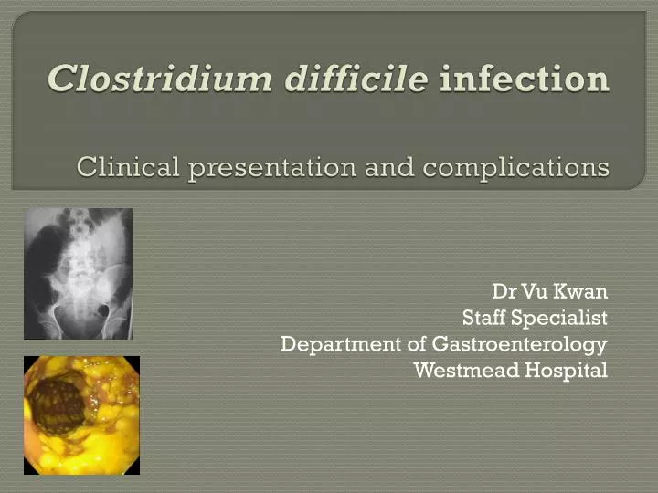
Clostridium difficileinfectionClinical presentation and complications Dr Vu Kwan Staff Specialist Department of Gastroenterology Westmead Hospital
Mr HL • 72 year old male • Background: • Ischaemic heart disease • NSTEMI 2009 • Coronary stent • Echocardiogram: EF 25% • Atrial fibrillation • Warfarin • Chronic kidney disease • Baseline creatinine ~180
October 2009 • Per rectum bleeding • Admitted for observation • Discharged for outpatient colonoscopy • Recurrent bleeding • Admitted for inpatient colonoscopy • Colonoscopy: • Multiple large colonic polyps • Endoscopic mucosal resection performed • Histology • Multiple tubular adenomas • Invasive malignancy not excluded
Post-polypectomy • Represented 3 days post-procedure with recurrent rectal bleeding • ED assessment: • “Post-polypectomy bleeding” • “Possible peptic ulcer bleeding” • Commenced on high dose proton-pump inhibitor infusion • Observed for several days bleeding cessation • Discharged home
Representation Represented 2 days later with bloody diarrhoea Up to 10 episodes per day Initially assumed to be ongoing post-polypectomy bleeding No stool tests performed
Colonoscopy Pseudomembranous colitis
History • No history of recent antibiotics • Only history: • Elderly male • Multiple co-morbidities • Repeated hospitalisations • Only new medication = PPI
Progress • Commenced on oral metronidazole • Ongoing fluid balance problems • Dehydration due to diarrhoea • Worsening renal function • Fluid therapy resulting in pulmonary oedema • Prolonged HDU admission with other medical complications • Eventual resolution of diarrhoea & discharge 3 weeks later
Overview One of the most common healthcare-associated infections Spectrum of disease ranging from asymptomatic carriage to fulminant colitis Commonly a result of antibiotic therapy due to alteration of normal gut flora
Overview Can occur without antibiotic use, importantly via nosocomial transmission Mortality rates of up to~25% reported, particularly in elderly1 1. Crogan et al, GeriatrNurs 2007
1. Asymptomatic carriage Approximately 20% of hospitalised patients are C. difficilecarriers Significant reservoir for disease transmission Contribution of host’s immune response is unclear
2. C.difficilediarrhoea • Watery diarrhoea • >3 times per day • >2 days duration • More severe cases • Up to 15 motions per day • Lower abdominal pain and cramping • Low grade fever • Leucocytosis • Onset may be during antibiotic therapy or 5-10 days after treatment • Can present up to 10 weeks after antibiotic cessation
3. C.difficile colitis • More significant illness than diarrhoea alone • Constitutional symptoms, fever, abdominal pain + watery diarrhoea • Colonoscopy: • Non-specific diffuse or patchy erythematous colitis
4. Pseudomembranous colitis • The classic manifestation of full-blown C.difficile colitis • Symptoms similar to, but often more severe than, colitis due to other causes • Unwell, WCC, hypoalbuminaemia • Colonoscopy: • Classical raised white/yellow plaques
5. Fulminant colitis • Severe manifestation affecting ~3% • Account for the most serious complications: • Perforation • Prolonged ileus • Toxic megacolon • Death • Clinical features of fever, leucocytosis, abdominal distension
Extracolonic manifestations Small bowel Bacteraemia Reactive arthritis Others
1. Small bowel • Particularly described in small bowel subjected to recent surgery • Inflammatory bowel disease post ileal-anal anastomosis • Pseudomembrane formation • May act as a reservoir for recurrent colonic infection?
2. Bacteraemia Uncommon Associated with high mortality rate1 May be more common in patients with underlying gastrointestinal diseases2 Daruwala et al, Clin Med Case Reports 2009 Libby et al, Int J Infect Dis 2009
3. Reactive arthritis • Polyarticular arthritis • Knee and wrist in 50% of cases • Onset average 11 days after diarrhoea1 • Prolonged illness : average 68 days to resolve2 Birnbaum et al, ClinRheumatol 2008 Jacobs et al, Medicine (Baltimore) 2001
4. Other extracolonic manifestations Cellultis Necrotisingfasciitis Osteomyelitis Prosthesis infection Intra-abdominal abscess Empyema etc
Risk factors • General risk factors • Long duration antibiotics • Multiple antibiotics • Nature of faecal flora • Production of requisite cytotoxins • Presence of host risk factors • Specific risk factors • Immunosuppressive drugs • Gastric acid suppression • Cancer chemotherapy with antibiotic properties
Host risk factors Advanced age Nasogastric tube Severe underlying illness Prolonged hospitalisation Enema therapy GI stimulants Stool softeners
Inflammatory bowel disease • Chronic, relapsing inflammatory disorders of the bowel of unknown aetiology • Ulcerative colitis • Crohn’s disease • Enteric infections account for ~10% of ‘relapses’ • C.difficile in about half • May mimic a relapse, OR trigger a true relapse
Inflammatory bowel disease Crucial that C.difficile is considered in the differential diagnosis of every ‘flare’ Otherwise inappropriate escalation of immunosuppression may result in severe infection High index of suspicion required as classical pseudomembranes don’t form in IBD Treatment is to REDUCE their usual immunosuppressive drugs
Gastric acid suppression • Gastric acid inhibits germination of ingested C.dificile spores • Therefore, medications lowering gastric acid could increase risk of C.difficile infection • Clinical data are conflicting
Imaging investigations Abdominal xray CT scan Colonoscopy
Abdominal xray • Important in patients who are unwell with C.difficile infection • Findings: • Ileus • Toxic megacolon • Perforation
CT scan • Diagnosis can often be made on CT alone • Several characteristic findings: • Gross bowel wall thickening • Luminal narrowing • Characteristic signs: • “Accordion sign” • “Target sign”
Colonoscopy • Pathognomonic appearance of pseudomembranes • Raised, white/yellow plaques • Up to 1/3 right-sided only, so full colonoscopy better than sigmoidoscopy • Biopsies reveal spectrum of mucosal inflammation and necrosis
Colonoscopy • Beware colonoscopy in unwell patients with ileus or megacolon • Risk of perforation • If clinical picture and stool tests are suggestive, minimal role for colonoscopy