Download
1 / 74
750 likes | 764 Views
Fundamentals of protein and nucleic acid structure. Lecture 2 Structural Bioinformatics Dr. Avraham Samson 81-871. Tree of Life. All known life forms use the same building blocks suggesting there was a common ancestor. The tree of life is not that simple!.

E N D
Fundamentals of protein and nucleic acid structure Lecture 2 Structural Bioinformatics Dr. Avraham Samson 81-871
Tree of Life All known life forms use the same building blocks suggesting there was a common ancestor
The tree of life is not that simple! Gene transfer across kingdoms continually occurs (cyanobacteria became chlorophylls, proteobacteria mitochondria, viruses)
29 atoms of life Most common elements:H, O, C, N, P, S (97% of organismweight) Most common ions: Ca2+, K+, Na+, Mg2+, Cl-
Forces affecting structure: • H-bonding • Van der Waals • Electrostatics • Hydrophobicity • Disulfide Bridges d 2.6 Å < d < 3.1Å 150° < θ < 180°
Forces affecting structure: • H-bonding • Van der Waals • Electrostatics • Hydrophobicity • Disulfide Bridges Repulsion דחייה Attraction משיכה d 3 Å < d < 4Å
Forces affecting structure: • H-bonding • Van der Waals • Electrostatics • Hydrophobicity • Disulfide Bridges d d = 2.8 Å “IONIC BOND” יוניקשר “SALT BRIDGE” מלחגשר E = Energy k = constant D = Dielectric constant (vacuum = 1; H2O = 80) q1 & q2 = electronic charges (Coulombs) r = distance (Å) Coulomb’s law
Forces affecting structure: • H-bonding • Van der Waals • Electrostatics • Hydrophobicity • Disulfide Bridges
Forces affecting structure: • H-bonding • Van der Waals • Electrostatics • Hydrophobicity • Disulfide Bridges Other names: cystine bridge disulfide bridge Hair contains lots of disulfide bonds which are broken and reformed by heat
Levels of protein structure Primary: Amino acid sequence Secondary: Local fold pattern of small subsequence Tertiary: Fold of entire protein chain Quaternary: Complex of multiple chains
Primary (1o)structure The amino acid sequence is the primary structure
Primary (1o)structure 20 amino acids
pKa = pH of 50% dissociation
Amino acid nomenclature Primary (1o)structure
Primary (1o)structure • Amino acids polymerize through peptide bonds to form polypeptides
Primary (1o)structure • N-terminal is the start of a polypeptide chain • Amino acids are also called residues
side chains צדדישייר שלד backbone Primary (1o)structure
Primary (1o)structure • Post translational modification are important because they can change the function of proteins (i.e. phosphorylation, acetylation, hydroxylation, carbohydrate and lipid modifications) N-terminal acetylation hydroxyproline O-phosphotyrosine γ-carboxyglutamate
Amino acids chirality Enantiomers –mirror images - תמונת ראי- אננטיומרים chiral center Cכיראלימרכז Dextro-Laevus in Latin
Most proteins: only L amino acids C chiral center is L configuration • איך לקבועקונפיגורציה L וD- • סדר את האטומים לפי עדיפותעלפיהכללים הבאים • כלל .1 אטום עם מספר אטומי יותר גבוה בעל סדר עדיפות יותר גבוה. • (I > Cl > O > N> C > H) • כלל .2 אם האטומים זהים, העדיפותעלפי האטומים המתמירים • (C(CH3)3 > CH(CH3)2 > CH2CH3 > CH3) • שים את האטום עם סדר העדיפות הכי נמוך מאחור. • קבע את כיווןהחץ , מסדר עדיפות הכי גבוה לככוון הכי נמוך. אם החץ: • עםכיוון השעוןקונפיגורציה D • נגד כיוון השעון קונפיגורציה L רמז יותר קל בשביל לזכור :) CORNתירס (
Torsion angles w, φ, and ψ • Unlike w, the two backbone dihedral angles φ and ψ are free to rotate • This rotation freedom allows protein folding Dihedral angles: -180o <φ < +180o -180o < ψ < +180o w is 0o or 180o w
Peptide bond is planar Cα, C, O, N, H, Cα all lie in the same plane
Torsion angle (w) is usually trans Steric hindrance Question: What other residues can be cis?
Except for X-Pro bond in which cis is preferred Steric hindrance Steric hindrance Steric hindrance allows both cis and trans (4:1 ratio)
Ramachandran Diagrams • Steric hindrance dictates torsion angle preference • Ramachandran plot show preferred regions of φ and ψ dihedral angles Preferred regions
Secondary (2o)structure Helices • a-helix • 310-helix • p-helix b Sheets • Antiparallel • Parallel Turns • b,g,a,and p Unstructured • α-helix is the most common • 3.6 residues per turn (number of residues in one full rotation of 360°) • 5.4 Å pitch (translation along axis for one full rotation of 360°) Hydrogen bond: i→i+4
Secondary (2o)structure Helices • a-helix • 310-helix • p-helix b Sheets • Antiparallel • Parallel Turns • b,g,a,and p Unstructured 310-helices are rare in proteins: 3.1 residues per turn (number of residues in one full rotation of 360°) 6 Å pitch (translation along axis for one full rotation of 360°) Hydrogen bond: i→i+3
Secondary (2o)structure Helices • a-helix • 310-helix • p-helix b Sheets • Antiparallel • Parallel Turns • b,g,a,and p Unstructured π-helices are rare in proteins 4.3 residues per turn (number of residues in one full rotation of 360°) 6 Å pitch (translation along axis for one full rotation of 360°) Hydrogen bond: i→i+5
Hydrogen bonding in helices • CO of residue (n) forms an h-bond with NH of residue: • (n+4) → α-helix • (n+3) → 310-helix • (n+5) → π-helix
Secondary (2o)structure Helices • a-helix • 310-helix • p-helix b Sheets • Antiparallel • Parallel Turns • b,g,a,and p Unstructured • In antiparallel b-sheets • Adjacent β-strands run in opposite directions • Hydrogen bonds (dashed lines) between NH and CO stabilize the structure • The side chains (in green) are above and below the sheet
Secondary (2o)structure Helices • a-helix • 310-helix • p-helix b Sheets • Antiparallel • Parallel Turns • b,g,a,and p Unstructured • In parallel b-sheets • Adjacent β-strands run in same direction • Hydrogen bonds (dashed lines) between NH and CO stabilize the structure • The side chains (in green) are above and below the sheet
Ribbon diagram of β sheet • In addition to being purely parallel or antiparallel, β sheets can be mixed, with strands running in both parallel and antiparallel directions • Arrow pointing to C-terminal end
Secondary (2o)structure CO of residue i forms h-bonds with NH of either residue i+2, i+3, i+5, or i+4 β-turn i→i+3 H-bond (most common) γ-turn: i→i+2 H-bond α-turn i→i+4 H-bond π-turn i→i+5 H-bond Helices • a-helix • 310-helix • p-helix b Sheets • Antiparallel • Parallel Turns • b,g,a,and p Unstructured
Secondary (2o)structure Helices • a-helix • 310-helix • p-helix b Sheets • Antiparallel • Parallel Turns • b,g,a,and p Unstructured
Tertiary (3o)structure • Proteins fold into compact tertiary structures with hydrophobic cores. (spacefill representation) Structural classification: http://www.cathdb.info/cgi-bin/cath/GotoCath.pl?link=class.html
Tertiary (3o)structure • It is a bit difficult to see and understand anything here (sticks representation)
Tertiary (3o)structure • To simplify representation, secondary structure diagrams are used
Quaternary (4o)structure Dimer of identical subunits (Cro protein of bacteriophage lambda)
Quaternary structure • Coat of rhinovirus • (common cold) • 60 copies of 4 • different subunits, • 3 outside, • red, blue, green
Protein structure databases • Primary: UniProthttp://www.uniprot.org/ • Secondary: DSSP http://rcsb.org • Tertiary: PDB http://rcsb.org • Quaternary: PQS http://www.ebi.ac.uk/pdbe/pqs/
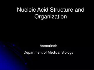
DNA and RNA Structure • NOTE: • Components • Sugar • Base • Phosphate • 5’ to 3’ direction • T->U in RNA • RNA - extra –OH at 2’ of pentose sugar • DNA - deoxyribose • Numbering • Single vs double strands • DNA more stable Voet, Donald and Judith G. Biochemistry. John Wiley & Sons, 1990, p. 792.
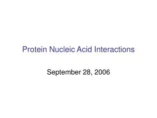
NOTE: • Pyrimadines and Purines • T->U in RNA • Names • Numbering • Bonding character • Position of hydrogen • Tautomers Purines The 5 Basesof DNA and RNA Pyrimadines