Download
1 / 39
390 likes | 395 Views
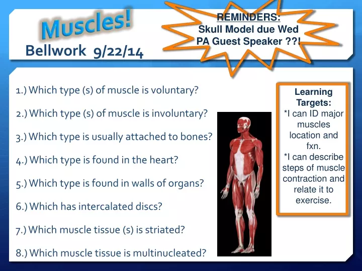
Muscles!. REMINDERS: Skull Model due Wed PA Guest Speaker ??!. Bellwork 9/22/14. 1.) Which type (s) of muscle is voluntary? 2.) Which type (s) of muscle is involuntary? 3.) Which type is usually attached to bones? 4.) Which type is found in the heart?

E N D
Muscles! REMINDERS: Skull Model due Wed PA Guest Speaker ??! Bellwork 9/22/14 1.) Which type (s) of muscle is voluntary? 2.) Which type (s) of muscle is involuntary? 3.) Which type is usually attached to bones? 4.) Which type is found in the heart? 5.) Which type is found in walls of organs? 6.) Which has intercalated discs? 7.) Which muscle tissue (s) is striated? 8.) Which muscle tissue is multinucleated? Learning Targets: *I can ID major muscles location and fxn. *I can describe steps of muscle contraction and relate it to exercise.
Muscles! REMINDERS: Skull Model due Wed PA Guest Speaker ??! Bellwork 9/23/14 Bellwork: compare 3 types of muscle (complete as much as possible) Classwork: labeling muscles and primary function Intro of sarcomere Learning Targets: *I can ID major muscles location and fxn. *I can describe steps of muscle contraction and relate it to exercise.
Muscles! REMINDERS: Skull Model due MONDAY (NO LATER) PA Guest Speaker ??! Bellwork 9/24/14 Bellwork: label and color skeletal muscle cell (on back of sarcomere sheet from yesterday) Classwork: sliding filament theory Learning Targets: *I can ID major muscles location and fxn. *I can describe steps of muscle contraction and relate it to exercise.
Muscles! Bellwork 9/29/14 Bellwork: 1. Sit with your partner, have skull and Self-evaluation completed! 2. review skeletal muscle structure and fxn Classwork: label and color skeletal muscle cell; I.D. fxns of all muscles. Learning Targets: *I can ID major muscles location and fxn. *I can describe steps of muscle contraction and relate it to exercise.
FUNCTIONS • Produce skeletal movement • Maintain posture and body position • Support soft tissues • Guard entrances and exits • Heat Generation body temp.
Types of Muscle Tissue • Skeletal • Cardiac • Smooth
SKELETAL MUSCLES • Includes all of the skeletal muscles that can be controlled voluntarily • Approx. 700 have beenidentified in the human body • Shape indicates function
Functional Characteristics of Muscle Tissue • Excitability: receive and response to stimuli • Contractibility: shorten forcibly when stimulated • Extensibility: stretch and extension • Elasticity: bounce back to original length after being shortened or lengthened.
TYPES OF MOVEMENTS BY MUSCLE CONTRACTIONS Each muscle begins at an origin, ends at an insertion, and contracts to produce an action. Origin: immovable (anchored end) Insertion: movable end of a muscle ** When a muscle contracts and shortens, its insertion is pulled toward its origin.
Types of Movement • Actions Include: • Flexion - a movement that reduces the angle between 2 bones; Opposite = Extension • Abduction - moving a part away from midline of body; Opposite = Adduction • Dorsiflexion - elevates top of foot and tilts it up; Opposite = Plantar flexion • Eversion – sole of foot out Opposite= Inversion • Supination - turn palm to anterior position; Opposite = Pronation • Rotation - movement around a longitudinal axis
Extension Flexion Dorsiflexion Extension Adduction Plantar Flexion Flexion Abduction
Supination Eversion Inversion Pronation Rotation
Muscle Organization Muscles are grouped according to their primary action: • Agonist- produces a particular movement (triceps) • Synergist- assists in producing movement • Antagonist - opposes agonist; relaxes during the prime movement
Connective Tissue Coverings • Epimysium: connective tissue that surrounds a skeletal muscle. • Perimysium: connective tissue that extends inward from the epimysium and separates the muscle into bundles of muscle fibers called fascicles. • Endomysium: Connective tissue that surrounds each fascicle.
Skeletal Muscle Structure • A muscle is composed of skeletal muscle tissue, nervous tissue, blood, and connective tissue. • Individual muscles are held in place by fascia, dense connective tissue. • Tendons are extensions of the fascia and intertwine with the fibers in the periosteum of the bone. • Muscles are connected to muscles by sheets of connective tissue called aponeuroses.
Organization of Muscle Fibers • Parallel- fascicles are parallel to the long axis of muscle (most muscles); strength • Convergent- fibers are based over a broad area, but all fibers come together at a common attachment site (pectoralis); more versatile in movement • Pennate- fascicles form a common angle with the tendon; uni, bi, multipennate; produces high tension • Circular/spinchter- fibers are concentrically arranged around an opening (orbicularis oris)
Skeletal Muscle Fibers • A muscle fiber is a multinucleated cell that attaches to connective tissue. • Sarcolemma is the muscle cell membrane. • Sarcoplasm is the cytoplasm containing nuclei, mitochondria, and myofibrils. • Myofibrils are composed of protein filaments, myosin and actin.
Protein Filaments • Myosin: Thick filament of twisted protein strands with globular ends called cross-bridges. • Actin: Thin filament protein, also contains tropomyosin and troponin. (movable) • Organization of filaments leads to light and dark striations seen in skeletal muscle.
Sarcomere Structure • Sarcomere: The fundamental Unit of a muscle; contains repeating patterns of striations • Striations: repeating light and dark regions
Sarcomere Structure • I bands (light bands) are composed of actin filaments attached to Z lines. • A bands (dark bands) are composed of myosin overlapping actin attached to Z lines by titin. • A central region (H zone) consists of myosin only with a thick line, the M line.
Microscopic structure • Sarcoplasmic reticulum: network of membranous sacs surrounding myofibrils. • Transverse tubules (T-tubules) extend into the sarcoplasm and contain extracellular fluid.
Microscopic structure • Cisternae: enlarged portions of the sarcoplasmic reticulum. • These three structures form a triad where the actin and myosin overlap.
http://highered.mcgraw-hill.com/sites/0072495855/student_view0/chapter10/animation__action_potentials_and_muscle_contraction.htmlhttp://highered.mcgraw-hill.com/sites/0072495855/student_view0/chapter10/animation__action_potentials_and_muscle_contraction.html • http://www.getbodysmart.com/ap/muscletissue/contraction/coupling/tutorial.html • http://www.getbodysmart.com/ap/muscletissue/contraction/multipleheads/tutorial.html
Muscle Contraction Quiz • Name of the fundamental unit of a muscle. • What are the two globular proteins (myofibrils) found in muscle? • What two proteins are associated with the thin filament? • What neurotransmitter cause stimulation of SR-sarcoplasmic reticulum? • What molecule is necessary for cross bridge formation? • When this molecule is released from the SR-sarcoplasmic reticulum, what does it bind to? • When a muscle contracts the length of the muscle ___________. • Describe a resting sarcomere.