Download
1 / 26
270 likes | 408 Views
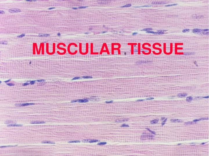
MUSCULAR TISSUE. Ⅰ . Introduction. 1. Composition: muscle cells ( muscle fibers ) + connective tissue . 2. Muscle fibers: elongated , containing myofilaments and being contractile . cell membrane → sarcolemma, cytoplasm → sarcoplasm .

E N D
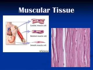
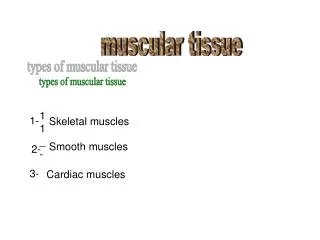
Ⅰ. Introduction 1. Composition:muscle cells (muscle fibers) + connective tissue. 2. Muscle fibers:elongated, containing myofilaments and being contractile. cell membrane →sarcolemma, cytoplasm →sarcoplasm. 3. Connective tissue:supporting, bring in blood vessels, lymphatics and nerves. 4. Classification: 3 types: skeletal, cardiac, and smooth muscle.
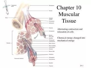
Ⅱ.Skeletal Muscle A. LM structure of Muscle fibers 1.Long, cylindrical cells 2.Multinucleated, with nuclei lying beneath sarcolemma 3. Having alternating dark and light bands (cross striations)
4. Myofibrils • Long, cylindrical in parallel • Aband, I band, Z line, H band, M line • Sarcomere: 1/2 I + A + 1/2 I ,shortest contractile unit of skeletal muscle.
B. Ultrastructure Myofibril Sarcoplasmic reticulum Mitochondrium Transverse tubule
a. Thick filaments • 1.5μm long and 10nm in diameter • Occupying A band • Made up of myosinmolecules: rods overlap; globular heads direct toward either of the ends forming cross bridgesandhave ATPase activity.
b. Thin filaments • 1μm long and 5 nm in diameter; • One end is inserted into the Z line, the other is free and extends into the A band; • Composed of actin, tropomyosin and troponin(TnI, TnT, TnC).
c. Arrangement • I band -- only thin filaments • A band -- both thick and thin filaments • H band -- only thick filaments • Z line -- anchor for thin filaments • M line –fixation of thick filaments
2. Transverse (T) tubules • Formed by sarcolemma invaginationat A-I junctions; • Form an anastomosing tubules encircling every myofibril; • Responsible for rapid conductionof impulses.
3. Sarcoplasmic reticulum (L tubule) • Network of smooth endoplasmic reticulum • Encircling each myofibril between 2 adjacent T tubules. • Ends dilated and fused to form terminal cisternae
T tubule + 2 terminal cisternae = a triad. • Storing Ca2+, regulating concentration of Ca2+ within sarcoplasm.
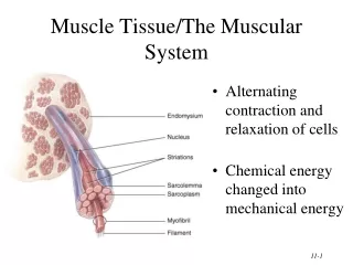
C.Muscles: made up of skeletal muscle fibers surrounded by CT. Ep F 1. Epimysium -- dense CT, surrounds entire muscle. 2. Perimysium -- fibrous sheath, surrounds muscle bundles. 3. Endomysium -- delicate loose CT, surrounds each muscle fibers. Pe Ed
III. Cardiac Muscle Pu A. General features 1. Found only in heart; 2. Has more connective tissue and capillaries; 3. Some specialized as Purkinje fibers.
B. LM structure 1. Short column in shape and branched; 2. show cross striations and fibrils, but less distinct; 3. Onecentrally placed nucleus;
C. EM structure 1. larger T tubules at Z line level; 2. Simple sarcoplasmic reticulum, small terminal cisternae; 3. Diadsare common consisting of T tubule and terminal cisternae on one side;
4. more sarcoplasm with more mitochondria and glycogen particles;
5.Intercalated discs • Specialized cell junctions at Z lines; • Longitudinal portion has gap junction providing synchronous contraction;
Transverse portion has desmosomes and fascia adherens to enhance intercellular junction.
Ⅳ. Smooth Muscle 1. Seen in blood vessels and hollow viscera, arranged in layers; 2. Spindle in shape, with an oval, centrally located nucleus;
3. Without striations, but contains thin and thick filaments; 4. Adjacent cells are linked by gap junctions.
Ⅴ. Mechanism of contraction -- sliding filament hypothesis 1. I band and sarcomere become shorter, H band shortens or disappears, and A band remains constant in length. 2. Thin filaments slide over thick filaments and insert further into the A band. 3. Ca2+ and ATP play an important role.