Download
1 / 11
110 likes | 118 Views
Lab Exercise 4:. Simple Staining and Bacterial Cell Morphology. Preparing a smear for staining. (The following procedure is used for all of our staining). 1. Flame (sterilize) your inoculating loop/needle before and after use. Heat from base to tip. Be sure to get the entire wire red hot.

E N D
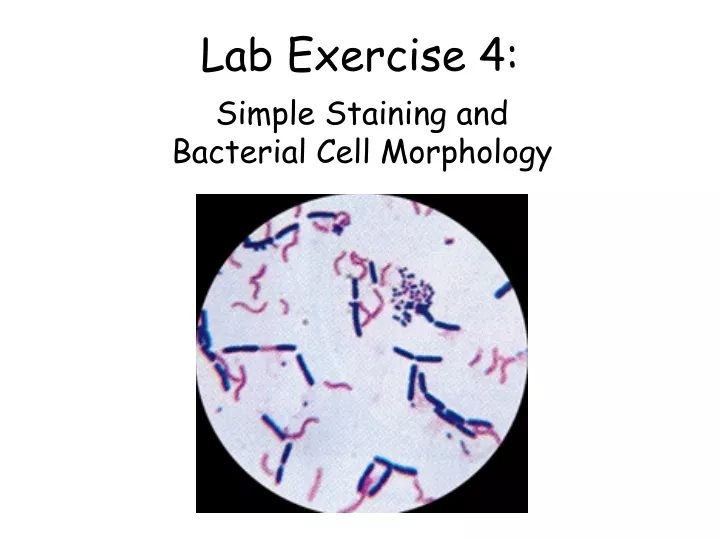
Lab Exercise 4: Simple Staining and Bacterial Cell Morphology
Preparing a smear for staining. (The following procedure is used for all of our staining) 1.Flame (sterilize) your inoculating loop/needle before and after use. Heat from base to tip. Be sure to get the entire wire red hot. Microbiology is Fun!
2. Prepare the smear a.If you have a solid culture (agar colony), place a small drop of distilled water on a clean slide. Drag the sterile inoculating needle tip through the edge of an isolated colony. b. Gently spread the mixture into a circle the size of a quarter. (A loop of liquid culture can be placed directly on the slide and spread out.)
3. Let the smear air dry completely. Do not apply heat while drying because this can lyse the cells.
4.Heat-Fix the smear. Smears are heat-fixed by quickly passing the slide through a flame (smear side up), two or three times. This causes the microbes to stick to the slide and not get washed off during the staining process.
5.Stain the smear. Place the slide on a staining rack over the sink. Flood the smear with stain and let it sit for 60-90 seconds. Rinse gently and blot dry.
6. Observe the slide under low and high-dry lenses to locate, center, and focus the image. Then, place a drop of oil directly on the stained smear (no cover slip). Turn the oil immersion lens into position and fine focus to observe the cells.
Name the bacterial morphologies (shapes and arrangements) seen here. 3 1 6 2 5 4 Answers: 1. Spirillum 2. Coccus 3. Bacillus 4. Diplobacillus 5. Streptobacillus 6. Diplococcus


![[Insert exercise name]](https://cdn0.slideserve.com/1400721/insert-exercise-name-dt.jpg)