Download
1 / 38
440 likes | 557 Views
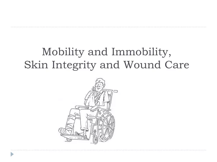
Mobility and Immobility, Skin Integrity and Wound Care. Mobility refers to a person's ability to move about freely. Immobility refers to: the inability to move about freely.

E N D
Mobility refers to a person's ability to move about freely. Immobility refers to: the inability to move about freely. NANDA definition of immobility: is a state in which the individual experiences or is at risk of experiencing limitation of physical movement.
Effect of immobility on physiological condition of clients including changes in the following systems • Metabolic. • Respiratory. • Cardiovascular. • Musculoskeletal. • Integumentary. • Urinary elimination.
Metabolic changes/ Deficiency of calories and protein causing Metabolic changes:Immobility disrupts normal metabolic functioning including: • Metabolic rate. • Metabolism of carbohydrates, fats, and protein. • Fluid and electrolyte imbalances. • Calcium imbalance. • GIT disturbances (anorexia, diarrhea, fecal impaction, and constipation). A deficiency of calories and protein causing: • Anorexia. • Negative nitrogen balance. • Weight loss. • Decreased muscle mass. • Weakness result from tissue catabolism. • Protein loss leads to decreased muscle mass, especially in the liver, heart, lungs, GIT, and immune system.
CHANGES Respiratory changes • Hypostatic pulmonary complications: • pneumonia • Leads to O2 , prolong recovery, and add to the client’s discomfort. • Decline in the client’s ability to cough productively. Increase of mucus distribution in the bronchi especially in supine, prone, or lateral position. Mucus accumulation in the airways. Because the mucus is an excellent media for bacterial growth hypostatic pneumonia result. Cardiovascular changes The three major changes are: • Orthostatic hypotension. • Increased cardiac workload. • Thrombus formation.
ORTHOSTATIC HYPOTENSION Orthostatic hypotension: is a drop of 25 mm Hg systolic and of 10 mm Hg diastolic in blood pressure when the client rises from a lying or sitting position to a standing position. Causes of Orthostatic hypotension in immobilization • Decreased circulating fluid volume. • Pooling of blood in the lower extremities. • These factors result in decreased venous return followed by a decrease in cardiac output which is reflected by a decreased in blood pressure increasing heart workload.
THROMBUS FORMATION: A thrombus: • is an accumulation of platelets, fibrin, clotting factors, and the cellular elements of the blood attached to the anterior wall of a vein or artery, sometimes occluding the lumen of the vessels. Factors that can cause thrombus formation: • Loss of integrity of the vessel wall (e.g., atherosclerosis). • Abnormalities of blood flow (e.g., slow blood flow in veins associated with bed rest and immobility). • Alterations in blood constituents (e.g., a change in clotting factors or increased platelet activity).
Musculoskeletal changes: Immobility lead to permanent impairment of mobility which causing: • Loss of endurance (staying power) of the muscles. • Decreased muscle mass. • Atrophy. • Decreased stability. • Impaired calcium metabolism. • Impaired joint mobility. Integrumentary changes: A pressure ulcer, or decubitus ulcer, is the consequence of ischemia and anoxia to tissue. Tissues are compressed, blood diverted, and blood vessels powerfully constricted by continual pressure on the skin and underlying structures; thus cellular respiration is impaired, and cells die.
Urinary elimination changes In the upright position, urine flows out of the renal pelvis and into the ureter and bladder because of gravitational forces. In recumbent or flat position, the kidneys and the ureters move toward a more flat surface. Urine format by the kidney must enter the bladder against gravity. Because the peristaltic contractions of the ureters are insufficient to overcome gravity, the renal pelvis may fill before urine enters the ureters (Urinary stasis). Urinary stasis increases the risk of: Urinary tract infection. Renal calculi. Renal calculus: Are calcium stones that lodge in the renal pelvis and pass through the ureters. Causes of renal calculi in immobilized client: Altered calcium metabolism. The resulting hypercalcemia
Predisposing factors with renal calculi formarion Fluid intake diminish. Other causes, such as fever. Increase the risk for dehydration. As a result of previous factors, urinary output declines on or about the fifth or sixth day. Urine become highly concentrated Causes of urinary tract infection: Concentrated urine. Poor perineal care after bowel elimination, particularly in women. Use of an indwelling urinary catheter.
Psychosocial effects of immobility: • Depression. • Behavioral changes. • Changes in the sleep-wake cycle. • Impaired coping.
Assessment clients for mobility: • Range of motion. • Gait. • Exercise and activity tolerance. • Body alignment: • Standing. • Sitting. • Lying.
The skin is the largest organ in the body and serves a variety of important functions in maintaing health and protecting the individual from injury. Impaired skin integrity is not a frequent problem for most healthy people but is a threat to older people, to clients with restricted mobility, chronic illnesses, or trauma, and those undergoing invasive health care procedures. Intact skin refers to the presence of normal skin and skin layers uninterrupted by wounds. The appearance of the skin and skin integrity are influenced by internal factors such as genetics, age, and the underlying health of the individuals as well as external factors such as activity.
A wound is a type of injury in which skin is torn, cut, or punctured (an open wound), or where blunt force trauma causes a contusion Body wounds are either intentional or unintentional. Intentional trauma occurs during therapy e.g., operations or venipuncture, removing tumor. Unintentional wounds are accidental; e.g. a person may fracture an arm in an automobile collision. If the tissues are traumatized without a break in the skin, the wound is closed. The wound is open when the skin or mucous membrane surface is broken.
Wounds may be described according to how they are acquired Incision wounds: Sharp instrument ''open, deep or shallow''. Contusion wounds: blow from a blunt instrument '' closed, skin appears ecchymosed (bruised)''. Puncture wounds: penetration of the skin and often the underlying tissues by a sharp instrument, either intentional or unintentional ''open wounds''. Lacerated wounds: tissue torn apart, often from accident ''open, edges are often jagged''. Abrasion wounds: Surface scrape ''open, involving the skin''. Penetrating wounds: penetrating the skin and underlying tissues. '' Open wound ''.
Types of wounds according to degree of wound contamination • Clean wounds: uninfected wounds in which minimal inflammation is encountered. • Clean – contaminated wounds: surgical wounds in which the respiratory, alimentary, genital or urinary tract has been entered. No evidenceof infection. • Contaminated wounds: open, fresh, accidental wounds and surgicalwounds involving a major break in sterile technique or a large amount of spillage from the gastrointestinal tract. Show evidence of inflammation. • Dirty or infected wounds: containing dead tissue and wounds with evidence of a clinical infection, such as purulent drainage.
Pressure Ulcers. Etiology of pressureulcers. Pressure Ulcers were previously called decubitus ulcers, pressure sores, or bedsores. It is any lesions caused by unrelieved pressure that result in damage to underlying tissues. Pressure ulcers are due to localized ischemia, a deficiency in the blood supply to the tissue. The tissue is compressed between two hard surfaces, usually the surface between the bed and the skeleton, when the blood cannot reach the tissue, the cells are deprived of oxygen and nutrients, waste products of metabolism accumulate in the cells, and the tissue consequently dies. Prolonged, unrelieved pressure also damages the small blood vessels. After the skin has been compressed, it appears pale, as if the blood had been squeezed out of it. When pressure is relieved, the skin takes on a bright red flush called reactive hyperthermia. The flush is due to vasodilatation, a process in which extra blood supply to compensate for the preceding period of impeded blood flow.
Risk factors Friction and Shearing Two other factors frequently act in conjunction with pressure to produce pressure ulcers: Friction: is a force acting parallel to the skin surface, such as sheets rubbing against skin create friction. Friction can abrade the skin, that is, remove the superficial layers, making it more prone to breakdown. Shearing force: combination of friction and pressure. It occurs commonly when the a client assumes a Fowler’s position. In this position, the body tends to slide downward toward the foot of the bed. This downward movement is transmitted to the sacral bone and the deep tissues . At the same time, the skin over the sacrum tends not to move because of the adherence between the skin and the bed linens. The skin and superficial tissues are thus relatively unmoving in relation to the bed surface, whereas the deeper tissues are firmly attached to the skeleton and move downward. This causes a shearing force in the area where the deeper tissues and the superficial tissues meet. and the superficial tissues meet. The force damages the blood vessels and tissues in this area.
Stages of pressure ulcers • Stage 1:- red color and the skin don’t return to normal color even thepressure is released. • Stage 2 :- redness accompanied by blisters or shallow break in the skin • Stage 3 :- break in the skin extending to the subcutaneous tissue • Stage 4:- ulcer involves loss of all skin layers exposing muscle and bone.
Wound Healing Healing is a quality of living tissue , it is also referred to as regeneration (renewal) of tissues. Healing can be considered in terms of types of healing and phases of healing. Types of Wound Healing There are two types of healing, influenced by the amount of tissue loss. 1- Primary intention healing Occurs where the tissue surfaces have been approximated (closed) and there is minimal or no tissue loss; it is characterized by the formation of minimal granulation tissue and scarring. It is also called primary union or first intention healing. e.g. closed surgical incision Primary intention healing is healing of a wound where the wound edges heal directly touching each other.
2- Secondary intention healing It is extensive and involves considerable tissue loss, and in which the edges cannot or should not be approximated. e.g., pressure ulcer. Secondary intention healing differs from primary intention healing in three ways: 1- The repair time is longer 2- Scarring is greater 3- Susceptibility to infection is greater
Phases of wound healing Inflammatory phase: is initiated immediately after injury and last 3 to 6 days. Two major processes occur during this phase: • Hemostasis • Phagocytosis Hemostasis (the cessation of bleeding) results from vasoconstriction of the larger blood vessels in the affected area, deposition of fibrin (connective tissue) and the formation of blood clots in the area. The blood clots, formed from blood platelets, provide a matrix of fibrin that becomes the framework for cell repair. The inflammatory phase also involves vascular and cellular responses to remove any foreign substances and dead and dying tissues. The area appears reddened and edematous. After 24 hours post injury, large macrophages enter the area these macrophages engulf microorganisms and cellular debris by a process known as phagocytosis.The macrophages also secrete angiogenesis factors which stimulate the formation of epithelial buds at the end of injured vessels, leads to reanastomosis. This phase include mildly elevated temperature, leukocytosis, and generalized malaise.
Proliferative phase: extends from day 3 or 4 to about day 21 postinjury. Fibroblasts (connective tissue cells), which migrate into the wound begin to synthesize collagen (whitish protein), these substance adds tensile strength, this decreases the chance that wound open again. Capillaries grow across the wound, ↑ the blood supply. Fibroblasts move from the bloodstream into wound, depositing fibrin , the tissue becomes a translucent red color. This tissue , called granulation tissues , is fragile and bleeds easily. Maturation(Remodeling phase): begins about day 21 and can extend 1 or 2 years after the injury. During maturation, the wound is remodeled and contracted. The scar becomes stronger but the repaired area is never as strong as the original tissue.
Types of wound exudate Exudate: - is material such as fluid and cells that has escaped from blood vessels during the inflammatory process and is deposited in tissue or on tissue surfaces. There are three major types of exudates:- 1- Serous exudate Consist chiefly of serum (the clear portion of the blood) derived from blood and the serous membranes of the body, such as the peritoneum. It looks watery and has few cells. e.g fluid in a blister from a burn.
2- Purulent exudate Is thicker than serous because of the presence of pus (leukocyte, dead tissue debris, dead and living bacteria). The process of pus formation is referred to as suppuration; bacteria that produce pus are called pyogenic bacteria. Purulent exudates vary in color, some acquiring tinges of blue, green, or yellow. The color may depend on the causative organism. 3- Sanguineous (hemorrhagic) exudates consist of large amounts of red blood cells, indicating damage to capillaries that is severe enough to allow the escape of red blood cells from plasma . Mixed types of exudates like: Serosanguineous ( consisting of clear and blood tinged drainage) purosanguineous (consisting of pus and blood )
Complications of wound healing 1- Hemorrhage Hemorrhage is abnormal massive bleeding; internal hemorrhage may be detected by swelling or distention in the wound. Hematoma, a localized collection of blood underneath the skin that may appear as a reddish blue swelling (bruise). The nurse should know the location of the pt’s incision to inspect the site of operation for bleeding at intervals for the first 48 hours, not less than Q 8hours. Any undue amount of bleeding should be reported, additional sterile dressing, fluid replacement, may need surgical interventions. Occurs in slipped sutures, dislodged clot, infection, erosion of blood vessels by a foreign
2- Infection Staphylococcus aurous, E. coli, Aerobacter aerogenes and pseudomonas aeroginosa. The main important area of prevention lies on aseptic techniques in wound care, cleanliness and environmental disinfection are important. The symptoms appear within 36-48 hours. The temperature and pulse increase, wound become tender, swollen, and warm. Nursing intervention will be through the use of warm antiseptic solutions to flush the wound. Take culture at site of operation. Specific antibiotics. A wound can be infected with microorganisms at the time of injury, during surgery, or postoperatively 3- Dehiscence with possible Evisceration Dehiscence: partial or total rupturing of sutured wound. Evisceration: the protrusion of the internal viscera through an incision area. A number of risk factors including obesity , malnutrition, multiple trauma, failure of suturing, coughing, vomiting, and straining, dehydration . Wound dehiscence is more likely to occur 4 to 5 days postoperatively. Sudden straining , such as coughing or sneezing, may precede dehiscence. The client may feel " something has given away “. When dehiscence or evisceration of a wound occurs, the wound should be supported by large sterile dressing moistures with sterile saline. Place the client in bed with knees bent to decrease pull on the incision. The surgeon is notified at once.