Download
1 / 44
440 likes | 446 Views
This course provides an in-depth study of the different structures of microorganisms, including capsules, filamentous structures, cell envelopes, cell walls, membranes, chromosomes, plasmids, ribosomes, and biological membranes. It covers the physiological activities that lead to these structures and their chemical composition. Offered by King Saud University, College of Science, Department of Botany and Microbiology.

E N D
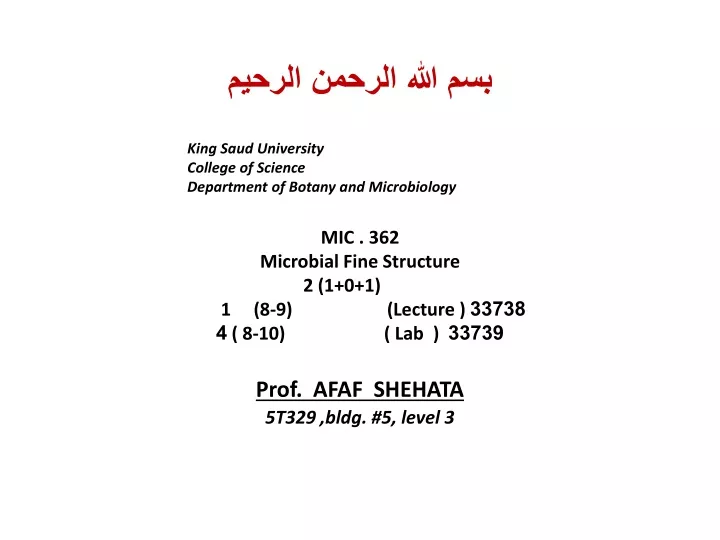
بسم الله الرحمن الرحيم • King Saud University • College of Science • Department of Botany and Microbiology • MIC . 362 • Microbial Fine Structure • 2 (1+0+1) • 1 (8-9) (Lecture ) 33738 • 33739 ( 8-10) ( Lab ) 4 • Prof. AFAF SHEHATA5T329 ,bldg. #5, level 3
362 حدق التركيب الدقيق للكائنات الحية الدقيقة 2(1+0+1) يعتبر المقرر النتيجة النهائية للعمليات الفسيولوجية حيث يستقرئ التراكيب المختلفة من حيث مكوناتها الكيميائية والوظيفة المناط بها .يدرس في هذا المقرر التراكيب التالية : العلبة- التراكيب الخيطية والزوائد- الغلاف الخلوي ويشمل الجدار الخلوي- الغشاء الخارجي والغشاء السيتوبلازمي- المورثات و الكروموسومات-البلازميدات الجراثيم- .الريبوسومات -الاغشية الحيوية - الكمون في الاحياء الدقيقة Microbial Fine Structure The courses outcome of the physiological activities so it does explain the resulting structures; The capsule - The filamentous structures -The cell envelope --The cell wall - The outer membrane - The cytoplasmic membrane - The genetic tools -The chromosomes - The plasmids - The spores - The ribosomes - The biological membranes . . MIC 362 Microbial Fine Structure Afaf Shehata
MIC 362 Lect. (1)LIVING ORGANISM A prokaryoteis a single-celled organism unicellular that lacks a membrane-bound nucleus (karyon), mitochondria, or any other membrane-bound organelles. Prokaryotes and Eukaryotes • Characteristics of Prokaryotes • First cells present for billions of years, were the only form of life on Earth. • Prokaryotes are the simplest type of cell. • Oldest type of cell appeared about four billion years ago. • Prokaryotes are the largest group of organisms • Prokaryotes unicellular organisms that are found in all environments. • Prokaryotes do not have a nuclear membrane . • Their circular shaped genetic material dispersed throughout cytoplasm. • Prokaryotes do not have membrane-bound organelles . • Prokaryotes have a simple internal structure. • Prokaryotes are smaller in size when compared to Eukaryotes.
A eukaryote is any organism whose cells contain a nucleus and other organelles enclosed within membranes. • Eukaryotes belong to the taxon Eukarya or Eukaryota. • Eukaryotic cells appeared on earth long after prokaryotic cells but they are much more advanced • Eukaryotic organisms unlike prokaryotic can be unicellular or multicellular . Eukaryotic • Characteristics of eukaryotes • Eukaryotic cells appeared approximately one billion years ago • Eukaryotes are generally more advanced than prokaryotes • Nuclear membrane surrounds linear genetic material (DNA) • Unlike prokaryotes, eukaryotes have several different parts. • Eukaryote’s organelles have coverings known as membranes. • Eukaryotes have a complex internal structure. • Eukaryotes are larger than prokaryotes in size .
Both types of cells have cell membranes (outer covering of the cell) • Both types of cells have ribosome's • Both types of cells have DNA • Both types of cells have a liquid environment known as the cytoplasm • Both have basic metabolism, like photosynthesis and reproduction. Similarities
MIC 362 Lect. (2) Structure of bacterial cell
Structure of bacterial cell Internal structures (Non-variant components or Essential components) External structures (Variant components or Non-Essential components) • Cytoplasmic membrane الغشاء البلازمى • Cytoplasm السيتوبلازم • Ribosome الريبوسوم • Mesosome الميزوسوم • Stored materials المواد المخزنة • Genetic material المادة النووية • Capsule or Slime layer العلبة • Cell wall الجدار الخلوى • Flagella الاسواط • Pili الاهداب
Primary Structure of Biological Macromolecules Determines FunctionProkaryotic structural components consist of macromolecules such as DNA, RNA, proteins, polysaccharides, phospholipids, or some combination .The macromolecules are made up of primary subunits such as nucleotides, amino acids and sugars (Table 1). It is the sequence in which the subunits are put together in the macromolecule, called the primary structure, that determines many of the properties that the macromolecule will have. Thus, the genetic code is determined by specific nucleotide base sequences in chromosomal DNA; the amino acid sequence in a protein determines the properties and function of the protein; and sequence of sugars in bacterial lipopolysaccharides determines unique cell wall properties for pathogens. The primary structure of a macromolecule will drive its function, and differences within the primary structure of biological macromolecules accounts for the immense diversity of life.
Prokaryotic Cell Architecture • A proKaryotic cell has five essential structural components: • a nucleoid (DNA) • ribosomes • cell membrane • cell wall • surface layer, which may or may not be an inherent part of the wall. • Structurally, there are three architectural regions: • 1- appendages (attachments to the cell surface: flagella and pili or fimbriae) • 2- cell envelope consisting of a capsule- cell wall and plasma membrane • 3- cytoplasmic region that contains the cell chromosome (DNA) and ribosomes and various sorts of inclusions (Figure 1). Prokaryotic Cell Architecture
Figure 1. Cutaway drawing of a typical bacterial cell illustrating structural components. See Table 2 below for chemical composition and function of the labeled components.
Table 2. Summary of characteristics of typical bacterial cell structures
Figure 2 . Electron micrograph of an ultra-thin section of a dividing pair of group A streptococci (20,000X). The cell surface fimbriae (fibrils) are evident. The bacterial cell wall is seen as the light staining region between the fibrils and the dark staining cell interior. Cell division in progress is indicated by the new septum formed between the two cells and by the indentation of the cell wall near the cell equator. The streptococcal cell diameter is equal to approximately one micron.
MIC 362 Lect. (3): External Structures (Variant components or Non-Essential components Appendages: flagella, fimbriae and pili Salmonella enterica. Salmonella is an enteric bacterium related to E. coli. The enterics are motile by means of peritrichous flagella.Flagella Flagella are filamentous protein structures attached to the cell surface that provide the movement for most motile prokaryotes. * Prokaryotic flagella are much thinner than eukaryotic flagella. * The diameter of a prokaryotic flagellum is about 20 nanometers. *The flagella filament is rotated by a motor apparatus in the plasma membrane allowing the cell to swim in fluid environments. * Bacterial flagella are powered by proton motive force (chemiosmotic potential) established on the bacterial membrane, rather than ATP hydrolysis which powers eukaryotic flagella. *About half of the bacilli and all of the spiral and curved bacteria are motile by means of flagella. *About 50 genes are required for flagella synthesis and function.
The flagella apparatus consists of several distinct proteins: • A system of rings embedded in the cell envelope (the basal body) • A hook-like structure near the cell surface • The flagella filament. • The innermost rings: the M and S rings, located in the plasma membrane, comprise the motor apparatus. • The outermost rings: the P and L rings, located in the periplasm and the outer membrane respectively, function as bushings to support the rod where it is joined to the hook of the filament on the cell surface. • As the M ring turns, powered by an influx of protons, the rotary motion is transferred to the filament which turns to propel the bacterium.
Figure 3. The ultrastructure of a bacterial flagellum . The flagellum of E. coli consists of three parts: filament, hook and basal body, all composed of different proteins. The basal body and hook anchor the whip-like filament to the cell surface. The basal body consists of four ring-shaped proteins stacked like donuts around a central rod in the cell envelope. The inner rings, associated with the plasma membrane, are the flagella powerhouse for activating the filament. The outer rings in the peptidoglycan and outer membrane are support rings or "bushings" for the rod. The filament rotates and contracts which propels and steers the cell during movement.
Distribution of flagella over the surface: flagella are either polar (one or more flagella arising from one or both poles of the cell) or peritrichous (lateral flagella distributed over the entire cell surface). Flagella distribution is a genetically-distinct trait that is occasionally used to characterize or distinguish bacteria. For example, among Gram-negative rods, Pseudomonas has polar flagella to distinguish them from enteric bacteria, which have peritrichous flagella. Figure 4. Different arrangements of bacterial flagella, occurs in half the bacilli and most of the spirilla. Flagella arrangements, which can be determined by staining and microscopic observation, may be a clue to the identity of a bacterium.
Flagella were proven to be organelles of bacterial motility by shearing them off (by mixing cells in a blender) and observing that the cells could no longer swim although they remained viable. As the flagella were re-grown and reached a critical length, swimming movement was restored to the cells. The flagella filament grows at its tip (by the deposition of new protein subunits) not at its base (like a hair). • Procaryotes are known to exhibit a variety of types of tactic behavior, i.e., the ability to move (swim) in response to environmental stimuli. For example, during chemotaxis a bacterium can sense the quality and quantity of certain chemicals in its environment and swim towards them (if they are useful nutrients) or away from them (if they are harmful substances). • Other types of tactic response in prokaryotes include: • phototaxis • aerotaxis • magnetotaxis. • The occurrence of tactic behavior provides evidence for the ecological (survival) advantage of flagella in bacteria and other prokaryotes.
Detecting Bacterial MotilitySince motility is a primary criterion for the diagnosis and identification of bacteria, several techniques have been developed to demonstrate bacterial motility, directly or indirectly.1. flagella stains outline flagella and show their pattern of distribution. If a bacterium possesses flagella, it is presumed to be motile.
Figure 5. Flagellar stains of three bacteria a. Bacillus cereus b. Vibrio cholerae c. Bacillus brevis. Flagellar distribution is occasionally used to differentiate between morphologically related bacteria. For example, among the Gram-negative motile rod-shaped bacteria, the enterics have peritrichous flagella while the pseudomonads have polar flagella.
2. motility test medium : A semisolid medium is inoculated with the bacteria in a straight-line stab with a needle. After incubation, if turbidity (cloudiness) due to bacterial growth can be observed away from the line of the stab.Julius Adlerexploited this observation during his studies of chemotaxis in E. coli: he prepared a gradient of glucose by allowing the sugar to diffuse into a semisolid medium from a central point in the medium. This established a concentration gradient of glucose along the radius of diffusion. When E. coli cells were seeded in the medium at the lowest concentration of glucose (along the edge of the circle), they swam up the gradient towards a higher concentration (the center of the circle), exhibiting their chemotactic response to swim towards a useful nutrient. Later, Adler developed a tracking microscope that could record and film the track that E. coli takes as it swims towards a chemotactic attractant or away from a chemotactic repellent. This led to an understanding of the mechanisms of bacterial chemotaxis.
Figure 6. Bacterial cultures grown in motility test medium. The tube on left is a non-motile organism; the tube on right is a motile organism. Motility test medium is a semi-soft medium that is inoculated with a straight needle. If the bacteria are motile, they will swim away from the line of inoculation in order to find nutrients, causing turbidity or cloudiness throughout the medium. If they are non-motile, they will only grow along the line of inoculation.
3. direct microscopic observation of living bacteria in a wet mount. Most unicellular bacteria, because of their small size, will shake back and forth in a wet mount. This is Brownian movement, due to random collisions between water molecules and bacterial cells. True motility is confirmed by observing the bacterium swim from one side of the microscope field to the other side.
A Desulfovibrio species. The bacterium is motile by means of a single polar flagellum.
Fimbriae and Pili • *Fimbriae and pili are short, hair-like structures on the surfaces of procaryotic cells. • *they are composed of protein. • *Fimbriae are shorter and stiffer than flagella, and slightly smaller in diameter. *fimbriae have nothing to do with bacterial movement. • *Fimbriae are very common in Gram-negative bacteria, but occur in some archaea and Gram-positive bacteria as well. • * Fimbriae are most often involved in adherence of bacteria to surfaces, substrates and other cells or tissues in nature. • * In E. coli, a specialized type of pilus, the F or sex pilus, apparently stabilizes mating bacteria during the process of conjugation.*Common pili (almost always called fimbriae) are usually involved in specific attachment of procaryotes to surfaces in nature. • * In medical situations, they are major determinants of bacterial virulence because they allow pathogens to attach to (colonize) tissues and/or to resist attack by phagocytic white blood cells. • For example: • pathogenic Neisseria gonorrhoeae adheres specifically to the human cervical or urethral epithelium by means of its fimbriae • enterotoxigenic strains of E. coli adhere to the mucosal epithelium of the intestine by means of specific fimbriae • The M-protein and associated fimbriae of Streptococcus pyogenes are involved in adherence and to resistance to engulfment by phagocytes.
Figure 8. Fimbriae and flagella on the surface of bacterial cells. Left: dividing Shigella enclosed in fimbriae. Right: dividing pair of Salmonella displaying both its peritrichous flagella and its fimbriae. The fimbriae are much shorter and slightly smaller in diameter than flagella. Both Shigella and Salmonella are enteric bacteria that cause different types of intestinal diarrheas. The bacteria can be differentiated by a motility test. Salmonella is motile; Shigella is nonmotile.
Lect 4. The Cell Envelope: capsules, cell walls and cell membranesthe cell envelope is a descriptive term for the several layers of material that envelope or enclose the protoplasm of the cell. The cell protoplasm (cytoplasm) is surrounded by - Plasma membrane - Cell wall -capsule. The cell wall is a layered structure in Gram-negative bacteria. All cells have a membrane, which is the essential and definitive characteristic of a "cell". Almost all procaryotes have a cell wall to prevent damage to the underlying protoplast. Outside the cell wall, foremost as a surface structure, may be a polysaccharide capsule or glycocalyx. MIC 362 Lect. (4) The Cell Envelope: capsules, cell walls and cell membranes
. Figure 9. Profiles of the cell envelope the Gram-positive and Gram-negative bacteria.
The Gram-positive wall: *is a uniformly thick layer external to the plasma membrane. * It is composed mainly of peptidoglycan (murein). The Gram-negative wall: * Cell wall thin and multilayered. * It consists of a relatively thin peptidoglycan sheet between the plasma membrane and a phospholipid-lipopolysaccharide outer membrane. *The space between the inner (plasma) and outer membranes (wherein the peptidoglycan resides) is called the periplasm.
Capsules Most procaryotes contain some sort of a polysaccharide layer outside of the cell wall polymer. In a general sense, this layer is called a capsule. * A true capsule is a discrete detectable layer of polysaccharides deposited outside the cell wall. *A less discrete structure or matrix which embeds the cells is a called a slime layer or a biofilm. *A type of capsule found in bacteria called a glycocalyx is a thin layer of tangled polysaccharide fibres which occurs on surface of cells growing in nature.
Figure 10. Bacterial capsules outlined by India ink viewed by light microscopy. This is a true capsule, a discrete layer of polysaccharide surrounding the cells. Sometimes bacterial cells are embedded more randomly in a polysaccharide matrix called a slime layer or biofilm. Polysaccharide films that may inevitably be present on the surfaces of bacterial cells, but which cannot be detected visually, are called glycocalyx.
Figure 11. Negative stain of Streptococcus pyogenes .The halo around the chain of cells is the hyaluronic acid capsule that surrounds the exterior of the bacteria
Capsules are generally composed of polysaccharide; rarely do they contain amino sugars or peptides (Table 4). Table 4. Chemical composition of some bacterial capsules
Capsules have several functions : • Like fimbriae, capsules, slime layers, and glycocalyx often mediate adherence of cells to surfaces. • Capsules also protect bacterial cells from engulfment by predatory protozoa or white blood cells (phagocytes). • protect from attack by antimicrobial agents of plant or animal origin. • Capsules in certain soil bacteria protect cells from perennial effects of drying or desiccation. • Capsular materials (e.g. dextrans) may be overproduced when bacteria are fed sugars to become reserves of carbohydrate for subsequent metabolism.
Figure 12. Colonies of Bacillus anthracis. The slimy or mucoid appearance of a bacterial colony is usually evidence of capsule production. The capsule is an essential determinant of virulence to the bacterium.
*Some bacteria produce slime materials to adhere and float themselves as colonial masses in their environments. * Other bacteria produce slime materials to attach themselves to a surface or substrate. * Bacteria may attach to surface, produce slime, divide and produce micro colonies within the slime layer, and construct a biofilm, which becomes an enriched and protected environment for themselves and other bacteria.
A classic example of biofilm construction in nature is: The formation of dental plaque mediated by the oral bacterium, Streptococcus mutans. • The bacteria adhere specifically to the pellicle of the tooth by means of a protein on the cell surface. • The bacteria grow and synthesize a dextran capsule which binds them to the enamel and forms a biofilm some 300-500 cells in thickness. • The bacteria are able to cleave sucrose (provided by the host diet) into glucose plus fructose. • The fructose is fermented as an energy source for bacterial growth. • The glucose is polymerized into an extracellular dextran polymer that cements the bacteria to tooth enamel and becomes the matrix of dental plaque. • The dextran slime can be depolymerized to glucose for use as a carbon source, resulting in production of lactic acid within the biofilm (plaque) that decalcifies the enamel and leads to dental caries or bacterial infection of the tooth.
Figure 13. (Left) Dental plaque revealed by a harmless red dye. Human dental plaque.
Another important characteristic of capsules : is their ability to block some step in the phagocytic process and thereby prevent bacterial cells from being engulfed or destroyed by phagocytes. • For example: • the primary determinant of virulence of the pathogen Streptococcus pneumoniae is its polysaccharide capsule, which prevents ingestion of pneumococci by macrophages. • Bacillus anthracis survives phagocytic engulfment because the lysosomal enzymes of the phagocyte cannot initiate an attack on the poly-D-glutamate capsule of the bacterium. • Pseudomonas aeruginosa, that construct a biofilm made of extracellular slime when colonizing tissues, are also resistant to phagocytes, which cannot penetrate the biofilm.