Download

1 / 6
60 likes | 115 Views
Supplemental Figure S1. A. MDA-MB-231 BCL-2. MCF-7 BCL-2. MDA-MB-231. MCF-7. Bcl-2. Bcl-2. Actin. Actin. B. Supplemental Figure S2. **. *. *. *. *. Control. - - - - - - -. Cisplatin 30 M.

E N D
Supplemental Figure S1 A MDA-MB-231 BCL-2 MCF-7 BCL-2 MDA-MB-231 MCF-7 Bcl-2 Bcl-2 Actin Actin B
Supplemental Figure S2 ** * * * * Control - - - - - - - Cisplatin 30 M - + + + + + + NAC 10 mM - - + - - - - TEMPO 2 mM - - - + - - - Tiron 10 mM - - - - + - - Trolox 200 M - - - - - + - U-74389G 5M - - - - - - +
Supplemental Figure S3 Cisplatin 30 M - + IP: Bcl-2 IB: 4-HNE IP: Bcl-2 IB: Bcl-2 MCF-7 Cisplatin 30 M - + IP: Bcl-2 IB: 4-HNE IP: Bcl-2 IB: Bcl-2 MCF-7 BCL-2
Supplemental Figure S4 IP: Bak Ab-2 IB: Bak Bak Bak Input IB: Bak MCF-7 BCL-2 Actin - - - - Control Cisplatin 30 M - + + + Trolox 200 M - - + - U-74389G 5M - - - +
Supplemental Figure S5 C A B
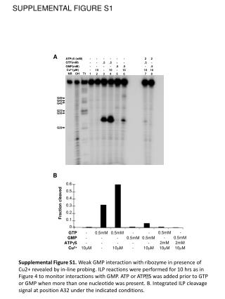
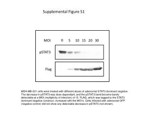
Supplementary Figure Legends Supplementary Figure S1. A, Immunoblot analysis of Bcl-2 expression in parental and Bcl-2-transfected MCF-7 and MDA-MB-231 cells. B, MCF-7 and MCF-7 Bcl-2 cells were treated with paclitaxel (20 nM), cisplatin (30 M) and HA14-1 (20 M) for 48 h and apoptosis was evaluated by M30 Apoptosense assay. Supplementary Figure S2. MCF-7 Bcl-2 cells with cisplatin (30 M) or cisplatin plus NAC (10 mM), TEMPO (2 mM), Tiron (10 mM), Trolox (200 M) or U-74389G (5 M) for 48 h. Apoptosis was detected by Annexin V staining. Columns, mean of three independent experiments; bars, SE. *, P < 0.05, **, P < 0.01. Supplementary Figure S3. MCF-7 and MCF-7 Bcl-2 cells were treated with cisplatin (30 M) for 12 h. (4-HNE)-histidine adduct formation was assessed by immunoprecipitation using anti-Bcl-2 (#2872) antibody followed by immunoblot analysis using anti-HNE monoclonal antibody. Immunoprecipitated proteins were also probed with anti-Bcl-2 antibody to test the efficiency of immunpoprecipitation experiments. Supplementary Figure S4. MCF-7 Bcl-2 cells were preincubated with lipid peroxidation inhibitors (Trolox, U-74389G) for 1 h and then treated with cisplatin for 36 h. Activation of Bak was assessed by immunoprecipitation using active conformation-specific anti-Bak (Ab-2) antibody followed by immunoblot analysis. 5% of the input for immunoprecipitation was also subjected to immunoblot analysis. Actin was used as a loading control. Supplementary Figure S5. MCF-7 Bcl-2 cells were transfected with Noxa siRNA or scramble siRNA. Cells were treated with cisplatin (30 M) for 48 h.A,apoptosis was evaluated by M30 Apoptosense ELISA.B,MMP loss was evaluated by flow cytometry using MitoTracker Red CMXRos. C, caspase-9 activation was determined by fluorometric caspase assay.