Download
1 / 15
170 likes | 294 Views
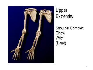
Upper Limbs 1 Fingers, hand, wrist. Dr Mohamed El Safwany, MD. Intended Learning Outcome. The student should be able to recognize technological principles of radiographic imaging of the upper limb. Pathological Indications (Upper limb).

E N D
Upper Limbs 1Fingers, hand, wrist Dr Mohamed El Safwany, MD.
Intended Learning Outcome The student should be able to recognize technological principles of radiographic imaging of the upper limb.
Pathological Indications (Upper limb) • FractureA break in the structure of bones. Types are : Simple, compound (open), incomplete (partial: includes the greenstick fracture), complete, comminuted, impacted, compression, depressed (ping-pong), pathologic, stress (fatigue), etc. Fracture names of the upper limb are (hand and forearm): Colles’, Pott’s, Barton’s, Smith’s,Bennett's, and Boxer’s. • Bursitis The inflammation of the bursa (fluid-filled sacs) enclosing the joints. • Carpal tunnel syndrome A common painful disorder of the wrist and hand due to a compression (by masses calcifications, ..) of the median nerve. • Colles’ fracture A transverse fracture of the distal radius, with distal fragment being displaced posteriorly, usually accompanied by an ulnar styloid fracture). • Smith’s fracture The reverse of Colles’ fracture (the fragment is displaced anteriorly).
Pathological Indications (Upper limb) • Rheumatoid arthritis Are inflammatory changes of the human body’s connective tissues (soft tissue swellings) around ulnar styloid process or MP joints. • Bone tumors May be benign or malignant (cancerous): Multiple myeloma : Is a primary cancerous bone tumor (most common). Osteochndroma : Is a benign type of bone tumor (most common). Osteosarcoma : Is the second most common type of malignant tumor. • Paget’s disease A chronic destructive skeletal bone disease leading to very dense (yet soft) bones that tend to fracture easily. Most commonly affects the pelvis, femur, the skull, the vertebra, clavicle, and the humerus. • Gout A form of arthritis. Blood uric acid is excessive, deposited on joints and/ or tissues. First attack is to the big toe (may also involve the thumb). • Joint effusion Accumulated synovial fluid or blood hemorrhage within a joint cavity due to a fracture, dislocation, soft-tissue damage, or could only be due to inflammation.
Pathological Indications (Upper limb) • Osteoarthritis Degenerative joint disease of gradual deterioration of articular cartilage with hypertrophic bone formation. It is generally part of the natural aging process. • Osteomyelitis Local/generalized bacterial infection of bone/bone marrow. May also be introduced by trauma or surgery. • OsteoporosisBone atrophy (reduction in quantity of the bone) of the skeletal bones in postmenopausal women and elderly men resulting in ‘thin’ bones. The affected bones usually tend to fracture easily.
Pathological Indications (Upper limb) • Chondromalacia patellae Is (Runner’s knee): The softening and/or wearing of cartilage under the patella at a later stage. • Ewing sarcoma A primary bone malignancy in children, mainly in diaphysis of the long bones. • Bone cysts Benign neoplastic bone lesions filled with a clear fluid near the knee joint in pediatric patients. • Osgood Schlatter's disease Is the inflammation of bone and cartilage in the anterior proximal tibia (tibial tuberosity) of children. • OsteoclastomaA(Giant cell tumor): A benign lesion in the proximal tibia/distal femur, usually affecting epiphyseal closure. • Osteogenic sarcoma Is a highly malignant primary bone tumor in long bones, usually causing gross destruction of the bone.
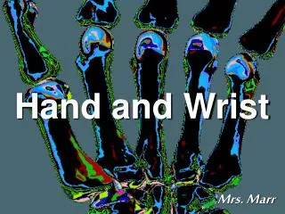
PA Fingers (Basic) • To show #s, dislocations, and pathology (osteoporosis, osteoarthritis, etc..) and any opaque foreign bodies (FBs). • Patient sits at the end of couch, elbow flexed 90, hand resting in pronantion on the film, all fingers extended and separated from each other. Sand bag over the lower forearm for immobilization. • Film: 18x24 cm. • CP: Proximal I.P.J. • CR: 90 vertically on the film center.
PA Fingers (Basic)
Lateral index finger (Basic) • To show #s, dislocations, and other pathology (osteoporosis, osteoarthritis, etc..) and foreign bodies (FBs). • Patient sits at the end of couch, hand medially rotated, the lateral aspect of index on the film, middle and other fingers flexed, sandbag over the lower forearm. • Film: 18x24 cm. • CP: Proximal I.P.J. • CR: 90 vertically on the film center.
PA Fingers (Basic)
AP thumb (Basic) • Shows fractures, dislocation, and pathology in distal and proximal phalanges. • Patient sits at end of the couch, shoulder at couch level, hand and wrist and forearm extended, arm internally rotated until posterior aspect of thumb rests supinated on the film, hand and wrist immobilized. • Film: 18x24 cm. • CP: First MPJ of the thumb. • CR: 90 to film center.
Assignment • Two students will be selected for assignment
Suggested Readings • Clark’s Radiographic technology
Question • Describe radiographic principles of hand radiogram?