Download
1 / 37
450 likes | 631 Views
MICROCIRCULATION – (CAPILLARY CIRCULATION). Dr.Mohammed Sharique Ahmed Quadri Asst Prof – Physiology Al Maarefa College. Objectives. Define capillary circulation. Recognize the importance of Mechanisms for control of blood flow to the various organs

E N D
MICROCIRCULATION – (CAPILLARY CIRCULATION) Dr.MohammedSharique Ahmed Quadri Asst Prof – Physiology Al Maarefa College
Objectives • Define capillary circulation. • Recognize the importance of Mechanisms for control of blood flow to the various organs • Understand factors affecting capillary circulation. • Identify Starling forces acting on capillary. • Appreciate the role of lymphatic. • Apply the physiological knowledge of microcirculation in understanding the causes of edema.
Arterioles • Major resistance vessels • Radius of arterioles supplying individual organs can be adjusted independently to • Distribute cardiac output among systemic organs, depending on body’s momentary needs • Help regulate arterial blood pressure
Arterioles • Mechanisms involved in adjusting arteriolar resistance • Vasoconstriction • Refers to narrowing of a vessel • Vasodilation • Refers to enlargement in circumference and radius of vessel • Results from relaxation of smooth muscle layer • Leads to decreased resistance and increased flow through that vessel
Mechanisms for control of arteriolar radius /blood flow to the various organs • Blood flow to organ depends on the demands of each organ. a) Local(intrinsic) control of blood flow is the primary mechanism that matches the blood flow to the metabolic needs of the organ (imp in determining distribution of cardiac out put). b) Neural or Hormonal(extrinsic) control is important in blood pressure regulation.
Local(intrinsic) controlof Arteriolar radius • Only blood supply to brain remains constant • Changes within other organs alter radius of vessels and adjust blood flow to organ • Local chemical influences on arteriolar radius • Local metabolic changes • Histamine release • Local physical influences on arteriolar radius • Local application of heat or cold • Chemical response to shear stress • Myogenic response to stretch
Local chemical influences on arteriolar radius • Following specific local metabolic factors are produce in the tissue as results of metabolic activity ,produces relaxation of arteriolar smooth muscle • Decreased O2 • Increased CO2 • Increased acid • Increased K+ • Increased osmolarity • Adenosine release • Prostaglandin release These factors produces relaxation od arteriolar smooth muscles by acting on vascular endothelium
Role of endothelial cells • Release chemical mediators(vasoactive substances) that play key role in locally regulating arteriolar caliber • Endothelial cells • Release locally acting chemical messengers in response to chemical changes as a result of metabolic activity in their environment. • Among best studied local vasoactive mediators is nitric oxide (NO), a potent vasodialotor
Local physical response to stretch Myogenic response to stretch Increase in blood flow Stretches the vessel Contraction of vascular smooth muscle Decrease blood flow back to normal
CONTROL OF REGIONAL BLOOD FLOW
LOCAL CONTROL OF BLOOD FLOW • Active Hyperemia - When any tissue becomes highly active [eg. Skeletal muscle during exercise]. the rate of blood flow through the tissue increases.(as a results of vasoactive metabolites) • When cells are metabolically more active they need more blood to bring in O2 and nutrients and remove metabolic waste. • Increase blood flow meets these increase local needs
LOCAL CONTROL OF BLOOD FLOW • Reactive hyperemia When blood flow to a tissue is blocked for few seconds and then is unblocked, the flow through tissue increases almost 4-7 times normal. • When blood flow to any region is blocked the arteriole in the region dilate due to • Myogenic relaxation in response to decrease stretch • Changes in local chemical composition ( similar to metabolically induced hyperemia) • After the occlusion is removed , blood flow to previously deprived tissue is transiently higher than normal due to widely dilated arterioles The excess blood flow lasts long enough to repay the tissue oxygen deficit that has occurred during occlusion.
LOCAL CONTROL OF BLOOD FLOW • Autoregulation – maintenance of constant blood flow to an organ in spite of fluctuations in BP. E.g. brain – auto regulation is best kidney – auto regulation is good skeletal muscle – auto regulation is poor • Two basic mechanisms that explain • Myogenic mechanism • Metabolic mechanism
Two basic mechanisms that explain local control of blood flow (auto regulation) : When MAP falls as in hemorrhage , blood flow to organs reduces 1. Myogenic mechanism decrease in blood flow Decrease Stretches on the vessel Relaxation of vascular smooth muscle Increase blood flow back to normal
Two basic mechanisms that explain local control of blood flow (auto regulation) : 2. Metabolic mechanism Decrease blood flow to organ Accumulation of vasodilator substances in active tissues Blood vessels dilate Increase blood flow Vasodilator metabolites O2 tension, H, CO2 tension, Temperature, K+, lactate, Adenosine, Histamine.
control of arteriolar radius /extrinsic control • Extrinsic control • Accomplished primarily by sympathetic nerve influence • Accomplished to lesser extent by hormonal influence over arteriolar smooth muscle • Epinephrin,norepinephrine, angiotensin II, vasopressin, etc.
SYMPATHETIC CONTROL OF ARTERIOLE • Sympathetic Control of Arterioles is important in regulating blood pressure. • Sympathetic ANS supplies arteriolar smooth muscle all over body except brain. • Increase sympathetic stimulation causes arteriolar vasoconstriction. • Decrease sympathetic stimulation causes arteriolar vasodilation. • There is no parasympathetic innervations to arterioles.
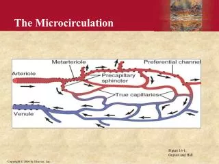
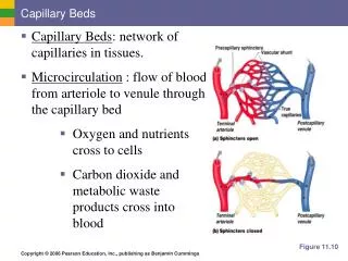
The Microcirculation • The term Microcirculation refers to the functions of the capillaries and the neighboring lymphatic vessels. • 5 % of circulating blood volume( 250 ml) is present in the capillaries at any given time. • This takes part into the exchange of nutrients, gases and waste products between the blood & tissues.
The Microcirculation • Over 10 billion capillaries with surface area of 500-700 square meters • Small volume of blood is exposed to larger surface area
Arteriole Meta arteriole Capillaries Venules. • Pre capillary sphincter is present at the junction where the capillary arises from the Meta arteriole. This opens and closes the entrance of capillary and hence regulates the blood flow through the capillary. • The capillary wall is thin & consists of a single layer of endothelial cells onbasement membrane. Pores are present between the endothelial cells that allow transport of substances including water.
Types of capillaries Classified according to the size of the pores • Brain – the pores are very tight and allow only very small molecules to pass thru. • Kidney & Intestine - the pores are wider – fenestrations • Liver - the endothelium is discontinuous with wide gaps between the cells.
EXCHANGE OF SUBSTANCES ACROSS THE CAPILLARY WALL This occurs by. • Simple diffusion Lipid soluble gases such as O2 & CO2 readily diffuse thru the endothelial cells. • Bulk Flow (various constituents of fluid moves in bulk/ unit ) - the most important mechanism for fluid transfer driven by Starlings forces. • A volume of protein free plasma filters out and reabsorbs
The rate of filtration at any point along the capillary depends on a balance of forces – STARLINGS FORCES. • 1. Capillary hydrostatic pressure Arterial end = 37 mmHg Venous end = 17 mmHg • 2. Plasma colloid osmotic pressure = 25 mmHg • 3. Interstitial hydrostatic pressure = 0 -1 • 4. Interstitial colloid osmotic pressure = 0 mmHg
Fluid movement = Kf [ (Pc – Pi) – ( Πc - Πi ) Kf = filtration coefficient
Arteriolar end fluid moves out into tissue spaces • Venous end fluid enters into capillaries. • Any decrease in plasma proteins (hypoproteinemia) or increase in capillary hydrostatic pressure (cardiac failure) causes edema. (abnormal increase in interstitial fluid volume ) • Histamines, bradykinin increases capillary permeability edema.
Starling’s forces across capillary wall Absorption Filtration
Lymphatic circulation • Lymphatic system is responsible for bringing the interstitial fluid to vascular compartment. • Normal 24 hrs lymph flow is 2-4 L • Lymphatic capillaries lie in interstitial fluid close to vascular capillaries ,these capillaries merge into large lymphatic vessels & eventually into largest vessel, thoracic duct which empties into large veins .
Lymphatic circulation • The interstitial fluid enter lymphatic capillaries through loose junctions between endothelial cells . • Lymph flow back to the thoracic duct is promoted by contraction of smooth muscle in wall of lymphatic vessels & contraction of surrounding skeletal muscle . • Failure of lymphatic drainage can lead to Edema .
What is Edema? • Accumulation of fluid beneath the skin or in a body cavity • Palpable swelling produced by expansion of the interstitial fluid volume
Causes of Edema • Increase capillary pressure • Increase vascular volume • Heart failure • Kidney disease • Pregnancy • Venous obstruction • Thrombosis • Liver disease • Decrease colloidal osmotic pressure • Increase loss of proteins • Nephrotic syndrome • Burns • Decrease production • Starvation, Malnutrition • Liver disease
Causes of Edema • Increase capillary permeability • Inflammation • Allergic reaction • Tissue injury • Malignancy • Obstruction of lymphatic flow • Surgical removal of lymph nodes • Malignant obstruction • Infection ( filariasis)
References • Human physiology by Lauralee Sherwood, seventh edition • Text book physiology by Guyton &Hall,11th edition • Text book of physiology by Linda .s contanzo,third edition