Download
1 / 32
340 likes | 522 Views
Lectures on Medical Biophysics Dept. of Biophysics, Medical faculty, Masaryk University in Brno. Biosignals and their processing Thermometry. What is a biosignal?.

E N D
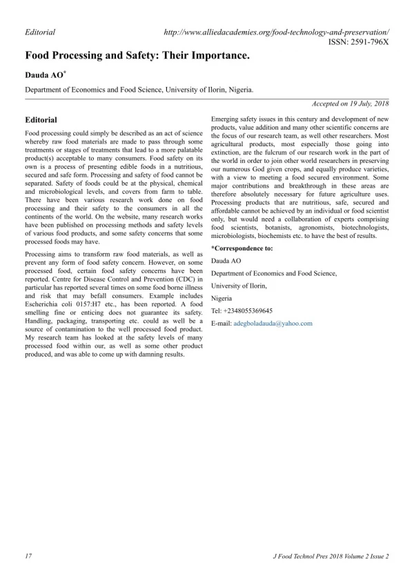
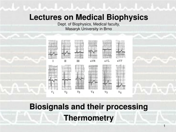
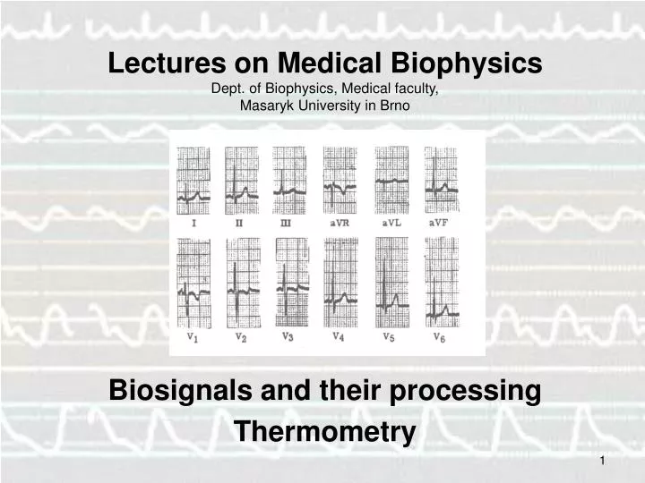
Lectures on Medical BiophysicsDept. of Biophysics, Medical faculty, Masaryk University in Brno Biosignals and their processing Thermometry
What is a biosignal? • Definition: a biosignal is a human body variable that can be measured and monitored and that can provide information on the health status of the individual. • Examples: • EKG (ECG): a V(t) biosignal which provides information on cardiac physiology / pathology • A US image: small voltage arising in elementary transducer by receiving reflection from tissue interface. • A CT tomogram: a m(x, y) biosignal for which the attenuation coefficient value is measured for each patient voxel at the position (x,y) in a slice of patient. • A 3-D MRI image: a SD (x,y,z) biosignal for which the hydrogen spin density (SD) is measured for each patient voxel at the position (x,y,z) in the patient each.
Types of Biosignals • ACTIVE (body generated) biosignals: the energy source for measurement derives from the patient himself (“internal source”) • Electrical active biosignals (known as BIOPOTENTIALS) e.g., EKG, EEG, EMG, ERG (electroretinogram) (ERG), EGG (electrogastrogram) etc • Non-electrical: e.g., temperature, blood pressure • PASSIVE (body modulated) biosignals: the energy source is from outside the patient (“external source) e.g., X-ray in CT • In this lecture we will be discussing active biosignals only
Origin of Biopotentials • Cells transport ions across their membrane leading to ion concentration differences and therefore charge differences - hence generating a voltage. • Most cell groups in the tissues of the human body do not produce electric voltages synchronously, but more or less randomly. Thus most tissues have a resultant voltage of zero as the various random voltages cancel out. • When many cells produce voltages synchronously the resultant voltage is high enough to be measurable e.g., EMG - muscle fibre contraction, most cells of the fibre perform the same electric activity synchronously and a measurable electric voltage appears.
Instruments for Measuring Active Biosignals • Biopotentials: Instrument consists of: • Electrodes: enable an electrical conductive connection between the examined body part with the measuring system • Signal processor (amplifier, ADC, electrical filters to remove noise, and unwanted frequencies etc) • Recorder (also called read-out device, today usually a computer monitor or a chart recorder) • Non-electric active biosignals: electrodes are replaced with appropriate sensors Two types of ECG electrodes Medical Temp sensors
Electrodes for Biopotentials: contact Voltage Problems • Problem: electrodes produce ‘contact voltages or contact potentials’ when put in contact with body! Polarisable electrodes producevariable contact voltage (via an electrochemical reaction) and hence are not suitable for accurate measurements. Non-polarisable electrodes produce a constant contact potential and hence are used when accurate measurements are required. Electrodes should be made of noble metals (metals which resist corrosion and oxidation). • Non-polarisable electrode: accurate measurements of biopotential. In practice, the silver-chloride (Ag-AgCl) electrode is most often used. • Polarisable: the contact voltage varies with movement of patient, humidity (sweating), chemical composition of ambient medium etc. • Concentration polarisation: the concentration of ions changes around electrodes due to electrochemical processes. • Chemical polarisation, gases are liberated on the surface of the electrodes.
Electrodes for Biopotentials: Electrode Sizes • Macro orMicroelectrodes. Latterused for biosignals from individual cells. Small tip diameter (<0.5 m) and made of metal (polarisable) or glass (non-polarisable). The glass microelectrode is a capillary with an open end filled with an electrolyte of standard concentration. • Superficial or needle electrodes. Superficialelectrodesare metallic plates of different shape and size. Good electric contact is ensured by a conducting gel. Their shape is often dish-like (see the Ag-AgCl electrode in the previous slide). Needle electrodesare used for recording of biopotentials from a small area of tissue. Used mainly for muscle biopotentials or long-term recording of heart or brain potentials.
Bipolar and Unipolar Electrode Pairs Bipolar electrode pair – both are placed in the electrically active region. Unipolar electrode pair, one electrode has a small area and is placed in the electrically active region. The second electrode (usually with a large area) is placed in an electrically inactive region (this electrode is called ‘indifferent’). A bipolar ECG electrode pair – depiction of the 1st limb lead
Signal processing: Amplifier • A high-fidelity (HiFi) amplifier is one which amplifies the biosignal without changing its shape (distortion). Modern medical must fulfil this condition. • Gain (amount of amplification) of an amplifier in dB = 20×log (Uo/Ui)
ECG - electrocardiogram Calibration 1mV voltage impulse • ECG (EKG) is the strongest and most often measured active biopotential. • In Europe 3 electrodes are placed on extremities (2 on arms, 1 on left leg), 6 electrodes are placed on chest. The right leg is used for an electrode which partially removes interfering voltages. • A pair of electrodes between which a voltage is measured, is called a lead. Every lead gives info on different parts of the heart.
Einthoven triangle Heart is modeled as a source of dipole electric field
2D and 3D Biopotential images Multiple electrodes placed on the surface of the body allow us to calculate voltage values throughout the torso (V (x,y,z) biosignal). Thus, we can localise problems with stimulus conduction throughout the myocardium.
EEG • -waves: f = 8-13 Hz, amplitude (A) max 50 V. Body and mind at rest. • -waves: f = 15 - 20 Hz, A = 5 - 10 V. Healthy people at full vigilance. • -waves: f = 4 - 7 Hz, A > 50 V. Physiological in children, in adults pathological. • -waves: f = 1 - 4 Hz, A = 100 V. Occurs in deep sleep under normal circumstances. In vigilance pathological. • In EEG record, some other patterns of electric activity can appear, characteristic of different brain diseases e.g., spike-wave complexes in epilepsy. • Brain biopotentials can be both spontaneous and evoked. Evoked potentials can be caused by sensory stimuli (vision, audition) or by direct stimulation by e.g. magnetic fields.
Anaesthesia: The EEG and the Bispectral Index • The Bispectral index monitor is a neurophysiological monitoring device which continually analyses a patient's electroencephalograms during general anaesthesia to assess the level of consciousness (too little anaesthetic and patient remembers, too much leading to brain damage). The essence of BIS is to take a complex signal (like the EEG), analyse it, and process the result into a single number which can be easily monitored. The BiS is the bottom trace.
The Bispectral Index is an example of a “descriptive indices”. These are not real physical quantities. They are parameters calculated from many measured parameters and by searching knowledge databases which contain measurements of many different patients (of various ethnic origins) with different health status.Complete algorithms of calculations and contents of knowledge databases are producer secrets. The medical doctorneeds only get acquainted with meaning of the respective index and the values which it can have,but it is not necessary to know how it is calculated. Comments on BiS etc.
... comments... It is usually enough to give some information about the patient for the computer to correctly search in the knowledge databases. It is almost always necessary to enter age, sex, race, body height and mass. There are sometimes strange questions about e.g. length of fingers or toes. Such “strange questions” are frequent ly found when monitoring the cardiovascular system. However, these questions can be important. When the respective answers are omitted, the software can use an incorrect statistical patient model and an incorrect index value will be displayed.
Artefacts • Definition: features of signals not arising from the target tissue • Arise from patient movement, electromagnetic waves in the environment (e.g., 50Hz electricity supply, mobile phones), patient movement, patient sweat etc
EKG Artefacts http://mauvila.com/ECG/ecg_artifact.htm 50Hz AC superimposed on the EKG Muscle tremors Moving baseline from patient movement, dirty electrodes, loose electrodes
Some EEG artefacts http://www.brown.edu/Departments/Clinical_Neurosciences/louis/artefct.html Pulse wave artefact: movement of electrode arising from patient pulse under the electrode. EKG signal artefact: EKG signal also picked up by the EEG electrodes. Both easily recognized because they are periodic.
Temperature Measurement “If a part of the human body is warmer or even colder than the surrounding parts, it is necessary to look for the disease focus in this place” Hippocrates
Main purposes of temperature measurements • monitoring of ill patients • monitoring of physiological reactions • monitoring of hyperthermia treatment Important specifications of thermometers: • accuracy • response time (determined by heat capacity of the sensor and its conductivity)
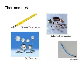
Types of Thermometry in diagnostics • 1. Point temperature measurement – measurement of temperature at individual points in the body • Contact • Dilation thermometers based on expansion (mercury and alcohol thermometers) • Digital thermometers based on thermistor sensors (resistance of thermistor changes with temperature) • Digital thermometers based on thermocouple sensors (voltage produced varies with temperature) • Contactless (ear tympanic thermometer) 2. Temperature distribution on the surface of the body (thermography) • Contact (use of sensors placed on skin) • Contactless – IR camera (other lecture)
Dilatation thermometers (i.e., based on expansion of some substance) Mercury-in-glass thermometer gives maximum temperature Its capillary is narrowed to avoid return of mercury into the reservoir. Disadvantage: long response time (long time necessary for a stable reading 3 - 5 min.) Medical high-speed thermometer: Alcohol filled – the capillary is not narrowed, the temperature must be read during the measurement, response time 1 min.
Tympanic (ear) thermometer Removable hygienic tip They are based on the measurement of infra-red radiation which is emitted from the ear drum. The temperature reading is obtained only 1 second after attachment of the sensor to the distal end of the acoustic meatus.
Physical principle of the temperature determination based on measurement of infrared radiation Stefan-Boltzmann law – dependence of the so-called spectral density of a black body radiation on temperature
Digital Thermometers: Thermistor sensor based R – resistance temperature in Kelvin T Ro – resistance at temperature To B – constant
Digital thermometers: Thermocouple sensor based Digital thermocouple sensor Thermovoltage U=a(t – t0)
Vojtěch Mornstein, Jan Dvořák, Věra Maryšková Last revision:January 2012 Authors: Presentationdesign: Lucie Mornsteinová Contentcollaboration and language revision: Carmel J. Caruana, Ivo Hrazdira