Download
1 / 59
600 likes | 738 Views
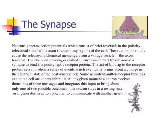
Synapse-to Nucleus Calcium Signalling. Why Calcium?. Na + and Cl - are sea water Excluded to maintain low osmotic pressure [K + ] i kept high for electrical neutrality [Ca 2+ ] i maintained very low Prevents precipitation of organic anions Mg 2+ helps solubilize organic anions.

E N D
Why Calcium? • Na+ and Cl- are sea water • Excluded to maintain low osmotic pressure • [K+]i kept high for electrical neutrality • [Ca2+]i maintained very low • Prevents precipitation of organic anions • Mg2+ helps solubilize organic anions
Calcium has been ‘selected’ by evolution as an intracellular messenger in preference to other monoatomic ions in the cell • Divalency - stronger protein binding than monovalent ions. • More flexible that smaller divalent Mg2+ ions more effective coordinate with protein-binding sites. • Energetically favourable to use Ca2+ as 2nd messenger (large [Ca2+] gradient) (10-7vs. 10-3M) – rel small amt needed to enter cell to incr signaling relatively little energy needed to pump it back out of the cell. • Higher [Ca2+] would ppt with PO43- ions lethal.
How cells keep [Ca]i low • All eukaryotic cells have PM Ca2+-ATPase • Excitable cells also have Na+/Ca2+ exchanger (NCX) • ER Ca2+-ATPase (against a high grad) • Mitochondrial high capacity (low affinity) pump • When [Ca]i very high (dangerous) levels (>10-5 M) • Inner mitochondrial membrane • Uses the electrochemical gradient generated during electron-transfer of oxidative-phosphorylation
Calcium Concentrations • [Ca2+]o / [Ca2+]i >104 • [Ca2+]o ~10-3 M • [Ca2+]ER ~10-3 M • [Ca2+]i <10-7 M at rest
Ca2+ - a versatile signal • Synaptic vesicle release (ms) • Excitation-contraction coupling (ms) • Smooth muscle relaxation (ms-sec) • Excitation-transcription coupling (min-h) • Gene transcription (h) • Fertilization (h)
How cells ↑ [Ca]i • Voltage-gated Ca2+ Channels • Membrane potential drives Ca2+ down its chemical gradient • Different channels in different cells • Different properties for different purposes
Ca2+ shut-off pathways • Voltage-gated Ca2+ channels inactivate • IP3 rapidly dephosphorylated • Ca2+ rapidly pumped out
Fertilization of an egg by a sperm triggering an increase in cytosolicCa2+ • 3 major types ofCa2+channels: • Voltage dependent Ca2+channels on plasma membrane • IP3-gated Ca2+release channels on ER membrane • Ryanodine receptor on ER membrane
Calcium uptake and deprivation • Na/Ca exchanger on plasma membrane, 2. Ca pump on ER membrane, 3. Ca binding molecules, 4. Ca pump on Mitochondia
Ca2+ as a 2nd Messenger (cont’d)Ca2+-Activated Signalling of Glu Receptor in the Postsynaptic Neuron
Synaptic Plasticity in the Nervous System • Activity-dependent plasticity is mediated by electrochemical activity of the synapse. • Activity-dependent plasticity is a change in neural connections and synaptic strength that are the hallmarks of learning and memory.
Targeting molecules for Calcium Calcium binding protein Calmodulin
Ca2+/calmodulin dependent protein kinase (CaM-kinase) Memory function: 1. calmodulin dissociate after 10 sec of low calcium level; 2. remain active after calmodulin dissociation
Ca2+/calmodulin dependent protein kinase (CaM-kinase) Frequency decoder of Calcium oscillation High frequence, CaM-kinase does not return to basal level before the second wave of activation starts
Synaptic plasticity in the Nervous System • Nervous system adapts to environmental changes. • Such stimulation activity-dependent plasticity or alterations in the number of synapses and/or in the strength of existing synapses.
The 3 Phases of Synaptic Plasticity • Early (sec-min) after electrical activity: changes in neural connections via modifications (phosphorylation) of existing proteins (ion channels) or delivery of proteins to postsynaptic membrane. • Intermediate (min-hr): synthesis of new proteins by existing levels of genes. • Late (days - longer ): changes in gene expression: txn and tln=> long-lasting changes. All of these phases triggered by Ca2+ influx.
Hippocampus – site of much plasticity and LTP studies. • Patients with hipp lesions anterograde and retrograde amnesia. • LTP – induced into postsynaptic neuron by high-freq. train of electrical impulses into presynaptic afferents. - model for learning and memory. - activity-dependent incr in synaptic efficacy that can last days-weeks in vivo.
LTP in the Hippocampus. • A model for plasticity - learning and memory. • Is an activity-dependent increase in synaptic efficiency that can last for days – weeks. • Induced in the postsynaptic neuron by repeated high-frequency stimulation of presynaptic afferents. • Characterized by an early, protein synthesis independent phase and late phases, which can be blocked by protein synthesis inhibitors. • During the longest phase, there is a critical period of transcription after the LTP-inducing stimuli has been applied. • Induction of LTP is critically dependent on an elevation of postsynaptic Ca2+.
IEGs – genes whose txn can be triggered without de novo protein synthesis (e.g., txn factors) 2ary wave of txn for other proteins required for LTP.
LTP in the Hippocampus (cont’d) e.g.,tissueplasminogen activator; activity-regulated cytoskeletal-assoc protein Ca2+ LTP-inducing stimuli IEGs zif268 c-fos c-jun NMDA receptors Secondary wave of txn, leading to the struct/func changes required for maintenance of LTP
Synaptic plasticity in the Nervous System – Control of Gene Expression • Pre-initiation complex. • Histoneacetylase activity. • RNA pol. • Transcription factors. • Promoter, enhancers, silencers. • REST/NRSF binding NRSE. • Signal-inducible transcription factors.
Control of Gene Expression • Control of gene expression can occur at any stage in the process. • By far, the most common point of regulation is at transcription initiation (RNA Pol II). • Transcription factors
Transcription Factors
Synaptic plasticity in the Nervous System – Ca2+-Responsive DNA Regulatory Elements and their Txn Factors • Cyclic-AMP response element (CRE). - Incr of synaptic activity synaptic NMDA receptor-dependent transient Ca2+ currents and long-lasting LTP in hipp (CA1 region) activate (phosphorylate) CREB txn factor CaMKII and MAPK (ERK) signalling pathways • Serum response element (SRE). - Induce expression of c-fospromotor activation of L-type Ca2+ channels. • Nuclear Factor of Activated T cells (NFAT) response element. - NFAT activity regulated by Ca2+-activated calcineurin. - Calcineurindephoscyto NFAT transport into nucleus. - W/o Ca2+-activated calcineurinactivity, NFAT becomes rephos by GSK and re-exported to cytoplasm.
Recall: Activating txn factors bind here, upstream, enhance the rate of PIC formation by contacting and recruiting the basal txn factors via adaptors or co-activators Txn factors can also acetylatehistones, disrupting/modifying chromatin structure Pre-initiation complex RNA Pol II Basal txn Core Promotor Element A wide variety of intracellular signaling pathways can influence the rate if txn initiation by many txn factors There are several well- characterized DNA elements that act as binding sites for txn factors that are regulated by Ca-activated signaling pathways Phosphorylation Reactions Stimulus
Ca-Responsive DNA Regulatory Elements and their Transcription Factors • cAMP-response element (CRE) – bound by CRE binding protein (CREB). - Ca activation of CREB is mediated by CaM KII and Ras-ERK1/2 signaling pathways.
Ca-Responsive DNA Regulatory Elements and their Transcription Factors • Serum Response Element (SRE) – binding site for serum-response factor (SRF) Ternary complex factor (TCF) TCF recognizes and binds SRE only with SRF bound Elk-1 SAP-1 SAP-2 SRF 5’ SRE Rsk 2 ERK 1/2 Ras Ca signaling pathways – dependent synaptic activation
Ca2+ as a 2nd Messenger (cont’d)Ca-Responsive DNA Regulatory Elements and their Transcription Factors • Nuclear Factor of Activated T cells (NFAT) Response Element Extracellular Intracellular Calcineurin NFAT-P Ca2+ NFAT P Cytoplasmic Nuclear NFAT NFAT-P Ca2+ Calcineurin (decr activity) GSK-3β ATP
Terminology: CRE(cyclic AMP response element); CREB: CRE binding protein; CBP: CREB binding protein
Physiological Importance of CREB • LTM • Information storage (Aplysia). • Confirmed by anti-sense oligonucleotides blocked LTM, but not STM formation. • Drug addiction. • Circadian rhythmicity. • Neuronal survival mediated by neurotrophins (BDNF). • Changes in synaptic strength and efficacy. • Besides BDNF, CREB-dependent pro-survival genes include nNOS, bcl-2, mcl-1 and VIP.
Mechanism of CREB Activation CREB Activation Requires a Crucial Phosphorylation Event • CREB binds CRE. • Ser 133. • Depol incr [Ca2+]cyto P-ser133 on CREB. • A133S abolished CREB-mediated gene expression of many IEGs. • CREB is a Ca2+-sensitive txn factor.
Mechanism of CREB Activation CREB Activation Requires a Crucial Phosphorylation Event Ca2+-dependent signaling molecules capable of phoshorylating CREB on ser133: CaMkinases and their role in Ca2+-activated, CRE-dependent gene expression: CaMKII, CaMKIV, and CaMKI. -Play roles in secretion, gene expression, LTP, cell cycle regulation, tln control. - Activate c-fos expression: - experiments with KN-62 decr L-type Ca2+ channel-activated c-fos expression. - experiments with calmodulin antagonist, calmidazolium. - CaMKIV – the prime member for CREB-mediate gene expression by nuclear Ca2+ signals. - experiments with anti-sense oligonucleotide disruption of CaMKIV expression abolished Ca2+-acticated CREB phosphorylation in hipp neurons. - critical for LT plasticity. - Knock-out mice for CaMKIV cognition/memory deficits related to noxious shock stimulus and related to spatial learning (hippocampus). - both inhibition of either CREB or CaMKIV function blocked cerebellar LTD (late phase) .