Download
1 / 17
170 likes | 261 Views
Chapter 4. Functional Anatomy of Prokaryotic and Eukaryotic Cells. Definition of “prokaryotic”. Refers to organisms, typically 1-celled, having cells which : lack a nucleus lack membrane-bound organelles contain 1 chromosome may contain extra-chromomal DNA (plasmids)

E N D
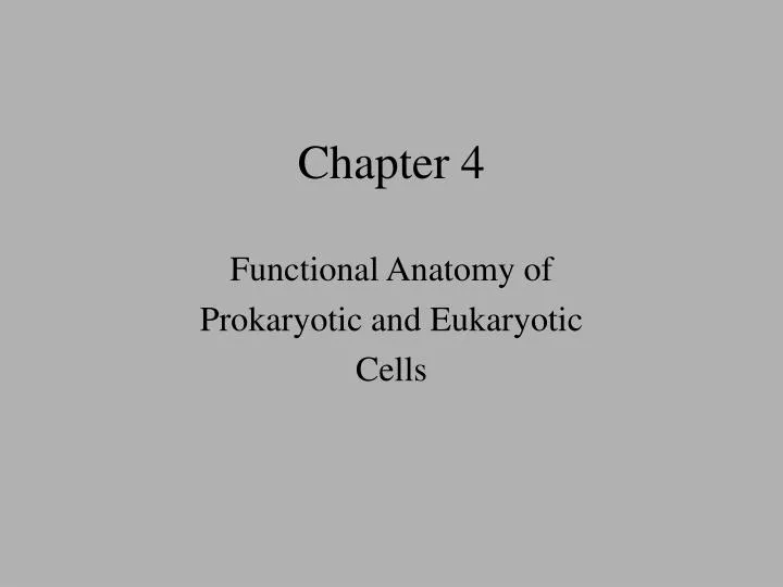
Chapter 4 Functional Anatomy of Prokaryotic and Eukaryotic Cells
Definition of “prokaryotic” Refers to organisms, typically 1-celled, having cells which: • lack a nucleus • lack membrane-bound organelles • contain 1 chromosome • may contain extra-chromomal DNA (plasmids) • contain 70S ribosomes • contain peptidoglycan cell walls
Bacterial cell size, shapes and arrangements • 2.0 – 10.0 uM in length • Eukaryotic cells ~10x larger • 3 common morphologies • bacillus = rod-shaped • coccus = spherical shaped • spirillum = spiral shaped • Many arrangements • diplo- • strepto- • Staphylo- ***spirochetes**
Bacterial morphologies • Morphology can be used as an initial identifier • However, shape can change in some bacteria depending on environs • “pleomorphic” cells
1) Cell wall structure • Alternating NAM & NAG amine sugars produce layers of block units • NAM = n-acetylmuramic acid • NAG = n-acetylglucosamine • Layers connected by tetrapeptide chains linked to NAM’s • Penta-glycine interbridges connect tetrapeptides in Gram + cells (sensitive to penicillin) • Direct peptide bonds connect tetrapeptides in Gram – cells (not sensitive to penicillin) Make up peptidoglycan
Gram positive cell wall structure Ok, not too bad – now for something completely different – Gram negative cell walls!
Gram negative cell wall structure Gram neg. cell walls are composed of peptidoglycan AND an outer membrane; it is multi-layered!!
Gram negative LPS* • *Lipopolysaccharide contains 3 parts: • Antigen O – can change shape in dif’t environs • Core polysaccharide – contains neg. charge • Lipid A – also called ‘endotoxin A’; released upon cell death and can have toxic affect on nearby cell membranes
Thick peptidoglycan 20-80 nm thick Retains CV-I complex of Gram stain Teichoic acid anchors cell wall to cell membrane and imparts a negative charge Glycerol-P polymer Two part structure Thin peptidoglycan (10-20 nm) Outer membrane Outer membrane contains LPS LPS imparts a negative charge Gram pos. vs Gram neg cell walls Gram + Gram -
2) Bacterial flagella • Composed of: 1) basal body, 2) filament, 3) hook • Basal body connects to cell wall and to cell membrane • Uses ATP to spin