Download

1 / 23
230 likes | 376 Views
龋病的临床表现. 西安交大医学院 侯铁舟 Tel 02988088507. 龋病的临床病理 一. 釉质龋 1. 釉质龋的组织学分区 透明层 暗层 损害体部 表层. 脱矿 再矿化. 2. 釉质龋的形成机理 3. 龋的再矿化过程 ? 釉质龋的早期发现方法. 脱矿 再矿化. 二.牙本质龋 1. 牙本质龋的组织学分区 感染层 (infected dentin)

E N D
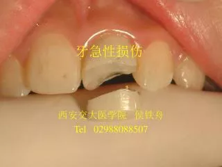
龋病的临床表现 西安交大医学院 侯铁舟 Tel 02988088507
龋病的临床病理 一. 釉质龋 1. 釉质龋的组织学分区 透明层 暗层 损害体部 表层 脱矿 再矿化
2.釉质龋的形成机理 3. 龋的再矿化过程 ? 釉质龋的早期发现方法 脱矿 再矿化
二.牙本质龋 1.牙本质龋的组织学分区 感染层(infected dentin) 影响层(affected dentin) 2.牙本质龋的形成机理
3.牙本质龋进展过程中牙本质及牙髓修复反应 牙本质牙髓复合体 再矿化 修复性牙本质
三.龋病病变过程特点 • 渐进性破坏 • 脱矿与再矿化交替 • 致病条件决定进展速度
临床特征.分类和诊断 一.龋病的好发部位 1.好发牙位 下颌>上颌, 后牙>前牙(第三磨牙除外) 2.好发牙面 窝沟>邻面>颊面(与滞留区有关)
二.分类和临床表现 1.按发病情况和进展速度分类 1)急性龋(acute caries) 猛性龋(rampant caries) 2)慢性龋(chronic caries) 静止龋(arrested caries) 3)继发龋(secondary caries)
2按损害的解剖部位分类 1)窝沟龋 2)根面龋 3)线性釉质龋(自学)
3. 按病变深度分类 1)浅龋 2)中龋 3)深龋
三. 龋病的诊断 1.龋病的检查方法 视诊,探诊,叩诊 温度试验 X线检查 牙线检查 透照法 荧光染色法
2.诊断标准 色,形,质的改变+症状
A traditional test of adequate caries removal during cavity preparation, is to hear the ring of a sharp probe on a hard dentine floor. Massler (1962) proposed a more conservative cavity preparation, which required the removal of infected dentine only, leaving behind soft but not infected, so called affected dentine. Affected dentine could be remineralised if the acid production was halted. Although Massler's ideas were supported by little experimental data, the idea of leaving behind soft dentine, and revisiting the tooth 6 weeks later, became an acceptable practice.