Download
1 / 32
340 likes | 636 Views
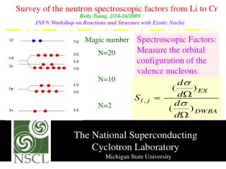
SPM Introduction Scott Peltier FMRI Laboratory University of Michigan. Slides adapted from T. Nichols. SPM!. Software to perform computation, manipulation and display of imaging data. SPM : Overview. Library of MATLAB and C functions Graphical user interface Four main components:

E N D
SPM Introduction Scott Peltier FMRI Laboratory University of Michigan Slides adapted from T. Nichols
SPM! Software to perform computation, manipulation and display of imaging data
SPM : Overview • Library of MATLAB and C functions • Graphical user interface • Four main components: • Preprocessing • Model Specification & Fitting • Inference & Results Interrogation • Supplemental Tools
2TR 3TR TR slice 1 slice 4 time SPM: Preprocessing • Eliminate systematic variation before statistical modeling • Slice timing • Adjust for variable acquisition time over slices • In UM processing stream, this is already done • “Realign”ment • Intrasubject registration • Motion correction • Done in UM stream
SPM: Preprocessing • “Coregister”ation • Intrasubject, intermodality registration • Registration of MR images with different TR/TE • Spatial “Normalize”ation • Intersubject registration • Register subject anatomy to atlas space SPM T1 template MNI space
SPM: Preprocessing • Spatial “Smooth”ing • Blur data into submission… • To satisfy random field theory assumptions • For intersubject analyses • “Segment”ation into GM/WM/CSF • Usually not directly used • Useful for structural studies Before convolution Convolved w/ circle Convolved w/ Gaussian Adapted from SPM course slides
SPM: Model Specification • “Specify 1st-level” • Specify the design, creating SPM.mat • “Specify 2nd-level” • T-tests (One or two sample, paired) • Regression • “Review” • Examine correlation of predictors • Power spectrum of experimental effects • “Estimate” • Fit a specified model based on a SPM.mat file
SPM: Inference • “ Results” button • First brings up “Contrast Manager” Can define single (t) or sets (F) of contrasts • Then displays MIP • MIP = Maximum Intensity Projection • Glass Brain • Can “surf” by dragging cursor
SPM: Inference • Interactive window • p-values • Correced for whole brain or subregion • Plotting of time courses • “Overlays” • Superimpose results on other images • Current location and value
SPM: Miscellaneous Tools • “Display” • Displays image with orthogonal sections • Check intensity values • Change origin • Change world space • i.e. Apply rotations/translations
SPM: Miscellaneous Tools • “Check Reg” • Display multiple images • Essential tool for assessing alignment of images • All images are displayed in the space of the first image
SPM: Miscellaneous Tools • “ImCalc” • Image calculator • Give one or more images, perform MATLAB arithmetic and write out result • “Utils” • Change directory • Results are written to current directory! • Delete files, etc.
SPM8 Batch Editor • Allows jobs to be saved, re-loaded, changed • Helps remove “Oops!” factor • Multiple steps can be loaded, run at once
SPM: Perspective • SPM tries to be a single solution for all fMRI processing and analysis, but there can be no such thing! • FMRI is a rapidly evolving field where each dataset has huge number of observations! • Don’t let SPM be a black box! • Understand what each component does • Understand how to get at the data • e.g. using ‘Display’, ‘Check Reg’
Resources • SPMweb site: http://www.fil.ion.ucl.ac.uk/spm/ • Introduction to SPM • SPM code download: SPM99, SPM2, SPM5, SPM8 • Documentation & Bibliography • SPM short course • Example data sets • SPM extensions • SPM email discussion list • Other software packages can complement SPM • MRIcron:http://www.mccauslandcenter.sc.edu/mricro/mricron/index.html • Quick and easy to read, display, and convert image data
Alternatives • FSL: http://www.fmrib.ox.ac.uk/fsl • Open source • Comprehensive tools for FMRI and DTI, has nice ICA analysis tool (MELODIC) • Free • AFNI: http://afni.nimh.nih.gov • Open source • Active community, multiple plugins • Free • BrainVoyager: http://www.brainvoyager.com • Excellent visualization • Closed source, ~$5k
SPM Spatial Transformations
Imaging data formats • Analyze format • .img Raw, binary data; 3D or 4D • .hdr Small binary header • Image dimension • Voxel size • NIFTI format • .img + .hdr • Like Analyze, but different .hdr definition • .niiSingle file! Header and Image file concatenated • World space transformation coded in NIFTI header
Is Left Right? Nose • Two conventions for viewing images • Neurological • On the screen, Left is Left side of subject • As if standing behind the head of the patient • Radiological • On the screen, Left is Right side of subject • As if standing at the foot of the patient • Standard in clinical radiology is, um, radiological • SPM always uses Neurological convention • Default for Analyze set by defaults.analyze.flip in spm defaults.m • flip = 0 ,Neuro., flip = 1 ,Rad. • NIFTI images allegedly have no ambiguity about left & right L R R L
Coregister & realignment • Coregistration & Realignment are rigid body transformations • Subject’s head doesn’t change size or warp between scans • Well, actually... • Each requires a “Reference” and a “Source” • Reference: Fixed image • Source: Image that is transformed • SPM modifies the .hdr file of the object image • Unless you explicitly ask it to, it doesn’t write out an image • Saves lots of disk space!
Voxel space vs. world space • Voxel Space • Just the original image • No reorientations or flips • World Space • Space defined by transformation from voxel to mm matrix M • Let v be a voxel location indexed from (1,1,1) • Then w=M*[v;1] is that location in world space, in mm • Can represent rotations, translations and flips
MNI Atlas Space Data Fresh from fMRI Lab Functional Space Functional images raprun_01.nii Low-res anatomy t1overlay.nii High-res anatomy t1spgr.nii Template image T1.niiscalped_avg152T1.nii
MNI Atlas Space Coregistration Functional Space Functional images raprun_01.nii Low-res anatomy t1overlay.nii High-res anatomy t1spgr.nii Reference Source CoregisterbuttonSets new world space in NIFTI headerDetermined from: Rigid body, M.I. registration of high-res to low-res anatomy Template image T1.niiscalped_avg152T1.nii
MNI Atlas Space After Coregistration Functional Space Functional images raprun_01.nii Low-res anatomy t1overlay.nii High-res anatomy t1spgr.nii (NIFTI header) Template image T1.niiscalped_avg152T1.nii
MNI Atlas Space Spatial Normalisation Functional Space Functional images raprun_01.nii Low-res anatomy t1overlay.nii High-res anatomy t1spgr.nii (NIFTI header) Normalizebutton Creates _sn.matfileDetermined from: Nonlinear, L.S. registration of high-res anatomy to T1 MNI template Template image T1.niiscalped_avg152T1.nii
MNI Atlas Space Spatial Normalisation Functional Space Functional images raprun_01.nii Low-res anatomy t1overlay.nii High-res anatomy t1spgr.nii (NIFTI header) _sn.matfile maps any Functional Space image to MNI space! Template image T1.niiscalped_avg152T1.nii
MNI Atlas Space After “Writing Normalized” Functional Space Functional Space Functional images raprun_01.nii Low-res anatomy t1overlay.nii High-res anatomy t1spgr.nii (NIFTI header) Template image T1.niiscalped_avg152T1.nii Normalized images wt1spgr.nii wraprun_01.nii
_sn.mat MNI Atlas Space Group Analysis: Strategy 1Only transform contrast img’s Functional Space rap_run’s beta’scon’sspmT’s Intrasubjectanalysis result Intrasubject analysis contrast images,transformed into atlas space (w/ _sn.mat),ready for group analysis wcon’s
Functional Space MNI Atlas Space _sn.mat Group Analysis: Strategy 2Transform all functionals wrap_run’s beta’scon’sspmT’s rap_img’s All functionaldata transformed into atlas space (w/ _sn.mat) Intrasubjectanalysis result con images ready for group analysis (already in atlas space)
Normalisation recommendations • With ‘scalped’ brains use ‘scalped’ template • Scalped template scalped_avg152T1.nii • Should give best results • We don’t care about scalp alignment! • Make sure WM equal in brightness • T1’s can have inhomogeneity artifact, where center of volume is brighter • Should apply homogeneity correction (bias correction) • UM: make sure to use het1spgr, het1overlay