Download
1 / 84
880 likes | 2.05k Views
Certification Review Course Peritoneal Dialysis Ray Agnello, BSN, RN, CNN Educator Saint Joseph’s Regional Medical Center Paterson, New Jersey. Objectives. To provide attendees with a summarized review of peritoneal dialysis

E N D
Certification Review CoursePeritoneal DialysisRay Agnello, BSN, RN, CNNEducatorSaint Joseph’s Regional Medical CenterPaterson, New Jersey
Objectives • To provide attendees with a summarized review of peritoneal dialysis • To highlight key points in the clinical care of a PD patient • Catheter Placement • Care of Catheter • Infectious Complication • Non Infectious Complications • Adequacy • Fluid Balance assessment of the PD patient.
Peritoneal Dialysis • Alternative to hemodialysis • Patient is taught to perform dialysis exchanges in the home setting • Focus is on patient autonomy and self care management • Patient must be followed by a licensed Peritoneal Dialysis unit & Nephrologist
Peritoneal Membrane • Translucent • Vascular membrane • Two layers Parietal (inner surface of abdominal wall) Receives blood supply from the arteries of the abdominal wall Visceral (covers abdominal viscera) Covers the abdominal organs Blood is carried by the mesenteric and celiac arteries Most vascular layer where most of the dialysis occurs Envelope of space between layers called peritoneal cavity Semi-permeable-acts as a Filter Kelley 2004
Anatomy and Physiology • Peritoneal Membrane • Semi-permeable • Bi-directional • Membrane size- 1-2 m2 • Vascular wall, interstitium, mesothelium , and adjacent fluid films • Closed in males • Women- ovaries and fallopian tubes open into the peritoneal cavity • Peritoneal cavity normally contains about 100 ml transudate
Kinetics of Peritoneal Dialysis • Diffusion • Osmosis • Ultrafiltration • Drug Transport
Diffusion Tea Bag = Peritoneal Membrane Water = PD Fluid Tea Leaves = Waste
Scheme of semi-permeable membrane:red = blood blue = PD fluidyellow = membrane .wikipedia.org/
Osmosis The diffusion of pure solvent across a membrane in response to a concentration gradient, usually from a solution of lesser to one of greater solute concentration. Miller-Keane 6th Edition
Osmotic Pressure of Dextrose Solution 1.5 % Solution 2.5 % Solution 4.25 % Solution
The Peritoneal Dialysis Process • Definition- intra (within) corporeal dialysis • Three Phases to the Exchange process • Drain • Fill • Dwell
How Does PD Work? • The semi-permeable peritoneal membrane lines the abdominal cavity and covers the abdominal viscera. • The membrane allows (via diffusion) the passage of toxins and electrolytes into the dialysis solution. • Ultra-filtration (removal of fluid) occurs via osmosis. • A “steady state” of toxin clearance and fluid management is achieved due to daily performance of dialysis. K. Kelly , RN NNJ Sept-Oct 2004
How Does PD Work? • Dialysis solution is infused and drained via a catheter that is surgically placed in the peritoneal cavity. • The action of draining and infusing dialysis solution is called an exchange. • The frequency of exchanges and volume is determined by the presence of residual renal function and the individual membrane characteristic.
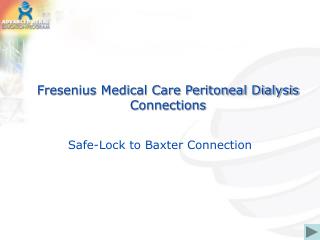
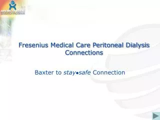
Infusion or Fill Baxter®
Drain Baxter®
Peritoneal Dialysis • Dialysis occurs during the dwell phase • Diffusion: solutes cross from area of greater concentration to lesser one -depends on concentration gradient -enough peritoneal surface area -size of fill volume • Ultra-filtration: water removal due to osmotic gradient between the hyperosmolar PD fluid and the capillary bed Kelley 2004
Historical Perspectives • Acute-Predominant use of PD prior to 1960’s • 1966- Automated cycler • 1967- Tenckhoff catheter • 1975- CAPD • 1978- Polyvinyl bags and manufactured in the US (prior PD fluid was available in glass bottles) • 1980’s- New catheter designs • 1987- PET and tidal PD -Twardowski • 1990’s-Alternative dialysate solutions, updated system designs ANNA Core Curriculum 5th Ed
Who Are the PD Patients ? • Choose PD as Renal Replacement Therapy • Hemodialysis Patient without Access • Failed allograft (transplanted kidney) • Have CHF or CVD which exempts them from hemodialysis • Often people with the benefit of CKD education
PD Patient Selection Inclusion Criteria Include Patients who: • Choose the modality • Want “control” • Prefer home for dialysis • Have residual renal function • CVD, CHF • Geriatric • Pediatric • Vascular Access Failure • Social support system available
Selection Continued Exclusion Criteria Patients who: • Have abdominal aortic aneurysm AAA (size dependent) • Derm. disease of the abdominal wall • Morbid abdominal obesity • Altered mental status, poor coping styles • Solitary life style • Patient states lack of interest in modality • Multiple abdominal surgeries- adhesions • Ostomies (increase risk of infection) • Recurrent hernias
Steps to PD Catheter Access • Evaluation by Nephrologist for PD catheter placement and identified as candidate. • Educated about catheter placement, pre and post operative care routines. • Referred to surgeon for evaluation that includes determination of exit site,clinical & anesthesia work-up, contraindications, completion of consent forms and scheduling of surgery.
Surgical EvaluationCatheter Insertion • Some units advocate insertion 2 to 6 weeks prior to dialysis to optimize healing. • Some units advocate insertion months in advance.(burying the catheter) • In most situations, PD access is elective
Surgical Evaluation • Abdominal wall weakness or hernia • Repair hernia preemptively or when symptomatic • Previous abdominal surgeries: multiple surgeries = increased likelihood of adhesions • Abdominal wall obesity
Pre Catheter Insertion • Patient Education and consent signed • Examination of the patient’s abdomen • Avoid scars and fat folds • Avoid beltline • Mark the abdomen • Surgical prep • Empty bladder • Patient showers with disinfectant soap • Bowel prep
Question Evidence-based practice suggests which of the following upon PD catheter implantation? • Large fill volumes immediately post-op • No need to wear a mask while performing PD exchanges • Incision site to be exposed to air during immediate post-op period • Administration of prophylactic IV antibiotics prior to catheter implantation to reduce the risk of peritonitis Core curriculum for Nephrology Nursing, 5th Edition. American Nephrology Nurses’ Association
Peri Operative RoutinesAnesthesia • Local infiltration with sedation • Intravenous propofol with Monitored Anesthesia Care • General anesthesia
Insertion Techniques • Bedside-temporary catheters • Laparoscopic placement • Surgical dissection • Buried Catheter technique • Percutaneous placement per Interventional Radiology
Insertion Techniques Buried catheter: • Entire catheter placed in subcutaneous pocket for 4-6 weeks or longer, allowing cuff & tunnel to heal • Exit site is externalized in a separate procedure • Reduced bacterial colonization(?) • Do not have long term outcomes yet Flanigan, Gokal, 2005
Catheter History • Early catheters were glass cannulas with straight or with mushroom ends • 1920-40’s: Various medical devices were used in the beginning of PD: needles, glass cannulas, sump drains, stainless steel coils, Foley catheters • 1923-Ganter used a needle for the 1st reported use in humans. • 1950’s-Nylon catheters, polyethylene, plastic with rounded tip & numerous tiny side holes • ANNA Core Curriculum 5th Ed
Catheter History • 1960’s- • silicon rubber catheters, with coiled intraperitoneal Tenckhoff & Schechter published results with silicone elastomer (Silastic ®) for chronic dialysis with 2 Dacron ® polyester felt cuffs • 1968-Tenckhoff cuffed straight catheter • 1970’s-single/double cuff coiled catheter; Toronto Western with 3 silicone disc • 1980’s-swan neck configuration (bent or curved SQ segment) Toronto Western with 2 silicone disc • 1990’s-t shaped catheter (Ash); Moncrief & Popovich technique for leaving the exterior segment buried SQ for 4 wk • The future..? • segment (Palmer, Quinton) • ANNA Core Curriculum 5th Ed
Catheters • Straight (single or double cuff) • Coiled (single or double cuff ) • Swan neck (single or double cuff) • Pre sternal swan neck • Toronto Western • Missouri catheters • Disc catheters
Cuffs • Single • Double • Elongated • Bead/flange configuration
Question… What is one advantage of implanting a cuffed PD catheter? • Acts as a barrier to prevent infection • Can only be used for CAPD • Ensures optimal adequacy • Can be implanted at the bedside Core curriculum for Nephrology Nursing, 5th Edition. American Nephrology Nurses’ Association
Adaptors • Plastic • Titanium
PD Catheter Access Complication • Immediate/Early Bloody effluent Pain with infusion Leak at exit site Exit site infection Migration of catheter tip Poor fill or drain, with or without pain Non-infectious cloudy effluent (lymphatic leak or eosinophilic peritonitis)
Question The patient’s fill volume is 2000mL. Upon draining, the patient’s volume is 1500mL. The nurse should assess the patient for which of the following? a. Peritonitis b. Catheter removal c. Constipation d. Subcutaneous tunnel infection Core curriculum for Nephrology Nursing, 5th Edition. American Nephrology Nurses’ Association
PD Catheter Access Complication • Later Issues • Exit site leaks or subcutaneous leaks • Pleural communications • Excessive granulation tissue • Chronic site or tunnel infection • Cuff extrusion • Cracked, brittle catheter • Repetitive episodes of peritonitis • Bowel perforations
Post Op • Follow up appointment with surgeon • Instructions (written & verbal) to patient, which include emergency contact numbers • Follow-up in PD unit within 48 to 72 hours of discharge • Pain medication/prescription • Reinforce dressing as needed • Teach patient to secure catheter • Flush catheter during training sessions
Remove primary dressing in 5 to 7 days by PD nurse • Dressing changed by PD nurse • Replace dressing with DSD, non-occlusive • Establish training schedule • Bowel regimen • No heavy lifting • Allow catheter to heal for 14 days or longer if possible before use Post Operative Discharge Plan Prevent Constipation
Peritoneal Dialysis Therapies • IPD (Intermittent Peritoneal Dialysis) • CAPD (Continuous Ambulatory Peritoneal Dialysis ) • CCPD (Continuous Cycling Peritoneal Dialysis) also known as APD (Automated Peritoneal Dialysis)
Training Sessions for the PD Patient • Assess readiness to learn • Provide a quiet, relaxed atmosphere for learning • Identify patient’s learning style • Individualized with respect to patient’s expectations, cultural beliefs, and coping abilities • Length of training based on patient’s clinical condition
On Call RN ON Call RN ON Call
Warming the Solution • Use warm, dry heat At home- PD heating pad • NEVER MICROWAVE!! Uneven heating of dextrose can create a 1st or 2nd degree burn to peritoneum Leaching of plastics into dialysate can Create a chemical peritonitis • NEVER MICROWAVE!!
Patients at risk for inadequate dialysis • No residual renal function • Low membrane permeability • Large patients • Patients not doing their treatments