Download
1 / 25
250 likes | 393 Views
The Cell Cycle & Cancer. Honors Bio. Human DNA Math for fun!. A single molecule of DNA has 1,000’s of genes! A single strand of DNA ~5cm long Each somatic cell has 46 molecules of DNA 5cm x 46 = 230 cm of DNA per cell! You have about 100 trillion cells

E N D
The Cell Cycle & Cancer Honors Bio
Human DNA Math for fun! • A single molecule of DNA has 1,000’s of genes! • A single strand of DNA ~5cm long • Each somatic cell has 46 molecules of DNA • 5cm x 46 = 230 cm of DNA per cell! • You have about 100 trillion cells • 230 cm x 100,000,000,000,000 = 23,000,000,000,000,000 cm of DNA in you body!!!!
How does it all fit? • SUPERCOILING!! • http://www.dnalc.org/resources/3d/07-how-dna-is-packaged-basic.html
Complete Mitosis POGIL Be sure to discuss each answer with your group!
Chromosome packaging • Eukaryotic chromosome – 1 d/s DNA molecule w/associated proteins • Each chromosome and its copy stay attached as sister chromatids and are attached by a kinetochore (protein) at the centromere (narrowing) • DNA winds twice around proteins called histones and is collectively referred to as a nucleosome • Why? • Prevents tangling • Regulates gene expression
Chromosomes • Most prokaryotic cells have 1 long circular chromosome • Number of chromosomes in the nucleus of eukaryotic cells varies by species • Genome– entire collection of genetic information within the cell • Multicellular organisms have 2 types of cells: • Somatic cells – all body cells • 2 sets of chromosomes (1 from each parent) • Gametes – reproductive cells (sperm & egg) • 1 set of chromosomes
Homework: • Find an article about the human genome project • Underline and define unfamiliar terms • Write a paragraph summary using the guidelines provided
Karyotype • Human Chromosome number • Somatic cells= Diploid (2n) 46 • Gametes = Haploid (n) 23 • Humans 23 pairs of homologous chromosomes • Autosomes 1-22 • Sex chromosomes 23 XX- female XY- male Preparation: • Using cells in tissue culture • Pretreating cells in a hypotonic solution, which swells them and spreads the chromosomes • Arresting mitosis in metaphase by a solution of colchicine • Squashing the preparation on the slide forcing the chromosomes into a single plane • Cutting up a photomicrograph and arranging the result into an indisputable karyogram.
Assignment • Select a Kayrotype to analyze • Analyze • Count Chromosomes • Determine Gender • Determine the chromosomal abnormality • Write a Letter to the parents • Detailing procedure of amniocentesis or chronic villi sampling • Description of a “normal” karyotype • Description of their child’s abnormal karyotpe • Possible reasons for the abnormality • Associated characteristics resulting from abnormality • 2 actual support groups available for the family • Any other pertinent information • Be sensitive as if you were actually contacting real parents
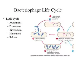
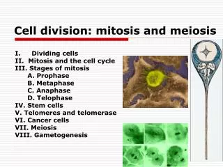
Cell Cycle • Cell Cycle: repeating sequence of cellular growth and division • G0 – non dividing cells • Interphase – growth and preparation • (90% of life): • G1 (1st growth phase) – cell grows and performs routine functions • S (Synthesis phase) – DNA is copied • G2 (2nd growth phase) – Microtubules are rearranged and cell prepares to divide • Mitosis – Nuclear Division • PMAT • Cytokinesis – division of cytoplasm
Interphase • G1 • Gap -Growth and normal function • S • Synthesis -Chromatin duplicated, not yet visible • G2 • Gap -2 centrosomesform • Animal cells – each centrosome features 2 centrioles (microtubules arranged in a circle)
Chromosome Duplication • During the S phase, each chromosome is duplicated via DNA replication • This results in 2 chromatids attached at thecentromere
Prophase • Chromatin condenses into visible chromosomes • Nucleolus disappears • Microtubule spindles form asters around centrosome • Microtubules move centrosomes to opposite poles of cell • Nuclear envelope fragments • Microtubules extend from centrosome to kinetochore of centromere
Metaphase • Centrosomes are at opposite ends of the cell • Chromosomes line up at midline or equator of cell • Each kinetochore has a microtubule that extends to the centrosome called a spindle fiber
Anaphase • 2 sister chromatids separate (becoming an individual chromosome) • Chromatids move to opposite poles as microtubules (spindles) shorten • Cell elongates • By the end, the two ends of the cell each have a identical set of chromosomes
Telophase • Nuclear envelopes and nucleoli reform • Chromosomes become less condensed and thread-like Animal Cells – cleavage furrow becomes visible
Cytokinesis - Animal Cells • Contractile ring mechanism -microfilaments called actin pinch the plasma membrane of the cell resulting in a Cleavage furrow
Cytokinesis - Plant Cells • No contractile ring, Why? • Cell walls stiff with cellulose • Vesicles (from golgi) fuse at midline to form cell plate • Cell wall forms from cell plate separating the 2 new plant cells
Challenge Question • Explain the structure and role of microtubules in animal cell division • Hint: MTOC Mitosis Cleavage Furrow • Pg 222-223 (cambell text)
Prokaryotic Cell Division • Binary Fission: divides into 2 new daughter cells • Also Asexual
Cell Cycle Control System • The timing and rate of cell division in various parts of an organism are crucial to normal growth, development, and maintenance • Molecules within the cell trigger and coordinate key events of the cell cycle • Checkpoint gene proteins can advance, delay or block the cell cycle • Both internal and external factors can affect cell cycling
Checkpoints • Checkpoint – critical control point where “stop” and “go” signals regulate the cycle (inspection station) • Regulation is influenced by environmental conditions • Stress (availability of nutrients) • Cell • 3 major checkpoints: • G1 (Most important in animal cells) • Is this a dividing cell? • Is this cell healthy? • Is this cell large enough? • **If cell does not receive the “go” signal, it will switch into G0 non-dividing phase • G2 • Is DNA copied properly? • M • Restarts cell cycle to G1 phase
Cancer • At the checkpoints • Oncogenes – tell cell cycle to “go” • Tumor Suppressor Genes – tell cell cycle to “stop” • If either of the genes that control the cell cycle become mutated • Mutagens – cause mutations • Carcinogens – mutagens cause cancer • The cell cycle goes too fast and divides out of control • Cells pile up in one area and form a tumor • Neoplasm such as a mole – benign tumor • Immune system does not recognize as foreign • Benign tumor – does not invade adjacent tissue • Malignant Tumor – cancer spreads and new tumors form (metastasize)
Malignant Cancer • Grow and develop abnormally • Cytoplasm shrinks and becomes disorganized • Plasma membrane becomes leaky (altered or missing proteins) • Metastasis – break away and establish new cell colonies in distant tissues • Can be lethal without treatment • Chemotherapy, radiation, surgery, or biotherapy