Download
1 / 28
420 likes | 1.08k Views
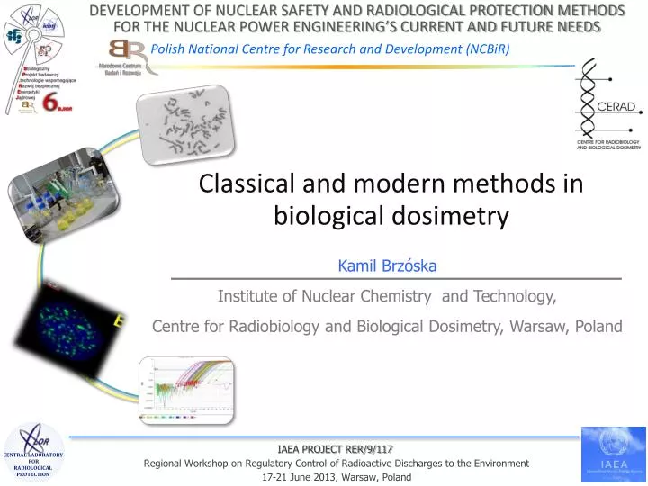
Classical and modern methods in biological dosimetry. Kamil Brzóska Institute of Nuclear Chemistry and Technology, Centre for Radiobiology and Biological Dosimetry , Warsaw , Poland. What is biological dosimetry ?.

E N D
Classical and modern methods in biological dosimetry Kamil Brzóska Institute of NuclearChemistry and Technology, Centre for Radiobiology and BiologicalDosimetry, Warsaw, Poland
What is biological dosimetry? Biological dosimetry is the quantification of exposure to ionising radiation by means of the measurable biological changes that take place in the system (biological indicators). Based on such indicators an individual’s exposure to ionising radiation can be detected and possible consequences of the exposure can be predicted. It enables suitable medical treatment to be planned when information from physical dosimetry is not available.
Whencan biological dosimetry be used? • When physical dosimetry information is not available as in the case of many accidents; • When the dosimetric information derived from physical dose meter is unreliable (e.g., non-uniform exposures); • Independently assess the damage as and when required for implementation of radiation protection standards; • To confirm suspected cases of overexposure; • Assessment of response in radiotherapy patients; • To check the compliance of dose limits for occupational exposures.
Characteristics of the ideal biological dosimeter • Should be sensitive in a wide dose range (20 mGy to several Gy); • Should show a reproducible dose response; • Should be a radiation specific; • Changes must occur early but remain stable for long time; • Should respond to all types of radiation (Low/high LET); • Partial body irradiations must be detectable and should enable the part exposed to be identified; • The technique should be as low invasive as possible; • It should be rapid and simple; • The technique should be amenable to automation.
Biologicaldosimetryassays • Blood cellscounts; • Dicentricchromosomalaberrationanalysis; • Micronucleusassay; • PrematureChromosomeCondensation(PCC); • Assay of stableaberrationsusingFluorescent in Situ Hybridization (FISH); • Histoneγ-H2AX foci formation assay; • Gene expressionanalysis; • Electron Spin Resonance (ESR) of dentalenamel; • skin speckle assaybased on radiation-inducedopticalchanges of skin;
Most biological dosimetry methods use peripheral blood lymphocytes • Blood sampling is a low-invasive procedure; • Lymphocytes circulate through the body and therefore even when only a part of the body was irradiated the dosimetry is possible; • They are synchronized and easy to culture. Peripheral human lymphocytes
Dicentric assay is a „gold standard” for biological dosimetry • Signal is detectable in lymphocytes, hence suitable even for partial body exposure; • Dicentrics are specific for radiation, their spontaneous frequency is very low in the healthy general population (about one dicentric per 1000 cells); • The frequency of dicentrics slowly decreases with time; • Realistic minimal detection level is about 0.2 Gy of whole body irradiation; • Maximum detection level is 5-8 Gy; • The method is reliable and most frequently used.
Mechanism of dicentric formation centromere acentric fragment chromosomalbreaks translocation dicentric
Dicentricassay centromers
Dicentircs dose-response curve for gamma Co60 radiation Dicentrics/100 mitoticcells Dose (Gy)
Disadvantages of the dicentric assay • Dicentric assay is time consuming and laborious, therefore not suitable for mass casualties scenarios; • The level of dicentrics in lymphocytes decreases with time, thus the retrospective dosimetry is unreliable; • To reveal dicentrics, lymphocytes must be induced to cell division using mitogen (e.g., phytohaemagglutinin) and cultured for 48 h before scoring can begin.
Objective 2: Development of biological dosimetry methods Phase 10: Evaluation of the relevance of combined PCC and FISH methods for biologicaldosimetry
Premature Chromosome Condensation (PCC)Assay • To visualize the alterations in DNA, chromosomes are artificially condensed using phosphatases inhibitors calyculin A or okadeic acid; • The method works only in the cycling cells, therefore lymphocytes have to bestimulated by phytohaemagglutinin (PHA); • Morphological changes such as: additional PCC fragments, PCC rings, dicentrics, translocations and unusually long chromosomes can be seen in G2/M cells (after 48h); • PCC method is usually used after very high doses of radiation, when the dicentric assay fails, because of cells stopped in cell cycle checkpoint G2/M;
Chemicallyinduced PCC after high doses of radiation Unusually long chromosomes PCC rings
RICA combines two methods Chromosome territories painting by FISH (Fluorescence In Situ Hybridisation) Chemically induced PCC in human lymphocytes in G0 phase of cellcycle + Okadeicacidorcalyculin A + ATP and CDK1
with PCC without PCC non-irradiated irradiated Prasanna et al. MutationResearch 466 (2000) 131-141
The advantages of RICA technique • It can be performed in unstimulated lymphocytes and therefore is faster than classical PCC, or dicentric assay; • High number of available cells; • It can be automated.
Results: • Two phosphatases inhibitors have already been tested: okadaic acid and calyculin A. Better results were obtain with okadeic acid, but still the number of condensed cells are to low to make any reasonable analysis; • In parallel to RICA we are validating classical chemically (calyculin A) induced PCC in G2/M lymphocytes; • Work in progress: creating the calibration curves in classical PCC for: PCC additional fragments, PCC rings and PCC unusually long chromosomes.
Objective 2: Development of biological dosimetry methods Phase 9: Evaluation of the relevance of gene expression analysis in blood cells for biologicaldosimetry
Geneexpression in bloodcells as a potentialbiologicaldosimeter Damageto macromolecules(DNA, proteins) Modification of activity of the cellularsignalingpathways Changes in geneexpression irradiation
Examples of the analyzed genes • Related to the DNA damage response: GADD45A, MDM2, DDB2; • Cell cycle control: CDKN1A, PLK3; • Programmed cell death (apoptosis): BAX, BCL2, BBC3; • Cellular response to stress: ATF3, SESN2, GDF15, FDXR; • Inflammatory response: TNFSF4;
Geneexpressionresults (1) TNFSF4 FDXR Examples of geneshighly but transientlyinducedafterirradiation.
Geneexpressionresults (2) DDB2 BAX Genes with stablyincreasedexpressioneven 48 h afterirradiation
Geneexpressionresults (3) BCL2 SESN2 Genesthatdidn’tprove to be deregulated by irradiation and thereforeare not suitable for biologicaldosimetry.
Preliminary conclusions from the gene expression analyses • Time after irradiation is the crucial factor in the analysis; • It is possible to identify irradiated samples; • Accurate dose prediction is difficult; • Interindividual variability is significant and must be taken into account.
Thank You for attention Prof. Marcin Kruszewski Prof. Anna Lankoff Dr Sylwester Sommer Dr Kamil Brzóska Dr Maria Wojewódzka Dr Teresa Bartłomiejczyk Iwona Buraczewska Tomasz Stępkowski Institute of Nuclear Chemistry and Technology, Centre for Radiobiology and Biological Dosimetry, Warsaw, Poland