Download
1 / 32
320 likes | 334 Views
What types of pathology can we identify and study from EKGs?. Arrhythmias Myocardial ischemia and infarction Pericarditis Chamber hypertrophy Electrolyte disturbances (i.e. hyperkalemia, hypokalemia) Drug toxicity (i.e. digoxin and drugs which prolong the QT interval). Anatomy.

E N D
What types of pathology can we identify and study from EKGs? • Arrhythmias • Myocardial ischemia and infarction • Pericarditis • Chamber hypertrophy • Electrolyte disturbances (i.e. hyperkalemia, hypokalemia) • Drug toxicity (i.e. digoxin and drugs which prolong the QT interval)
PR QRS AH HV How does the heart work
AV node activated by Atrial depolarizationSends signal through His-purkinje bundleGet depolarization of SEPTUM Left and Right BUNDLES transmit signal to Left and Right VENTRICLESNet “Vector” towards the LVShould be narrow (<120msec) if bundles working properlyThen have REPOLARIZATION = TwaveThe appearance of this electrical activity depends on which lead you are using to look at it
How to Look at an ECG • Rate: Is the heart rate too fast or slow? • Rhythm: Sinus rhythm or not? • Axis: Where does the majority of electrical activity point? • P wave: How big are the atria? • PR interval: How healthy is the AV node? • QRS wave: Is there abnormal conduction or a ventricular source? • QT: Long is bad • Ischemia and hypertrophy
ECG PaperCan Determine Heart Rate Rule: 300, 150, 100, 75, 60, 50 counting over for each big sqaure
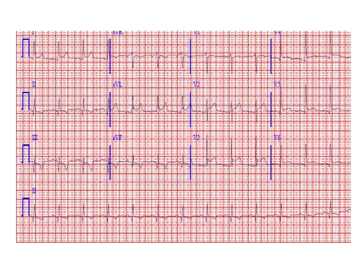
What is the heart rate? Answer = 75 per min
Rhythm : Is there a p wave? = Sinus Is it followed by a QRS?
Irregular pacemaker Multifocal atrial rhythm Atrial fibrillation Atrial fib/flutter Ectopic beats PVC PAC PJC Irregular conduction AV node block 1st degree: PR interval > 200 msec 2nd degree: Type 1: Wenkebach Type 2: dropped beat 3rd degree: p waves marching independent to QRS Reasons to have an irregular rhythm
Examples of Rhythms Multifocal Atrial Rhythm AFIB Atrial Flutter AFIB V TACH
EKG Leads 3 Standard Limb Leads 3 Augmented Limb Leads 6 Precordial Leads The standard EKG has 12 leads: The axis of a particular lead represents the viewpoint from which it looks at the heart.
The QRS QRS < 120 msec QRS > 120 msec Rabbit ears in V1 & V2 Wide S wave in V5 & V6 R axis deviation QRS > 120 msec Deep slurred S wave in V1 Wide R wave in V6, I & avL L axis deviation
Review • Rate: Is the heart rate too fast or slow? • Rhythm: Sinus rhythm or not? • Axis: Where does the majority of electrical activity point? • P wave: How big are the atria? • PR interval: How healthy is the AV node? • QRS wave: Is there abnormal conduction or a ventricular source? • QT: Long is bad • Ischemia and hypertrophy