Download

1 / 69
1.74k likes | 5.43k Views
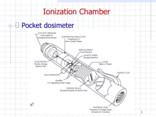
Ionization Chamber. Pocket dosimeter. Ionization Chamber. Pocket dosimeter. Radiation Quantities and Units. Radiation measurements require specification of the radiation field at various points At the source – Activity, mA, kVp In flight – Exposure, fluence (dN/dA), energy fluence (dE/dA)

E N D
Ionization Chamber • Pocket dosimeter
Ionization Chamber • Pocket dosimeter
Radiation Quantities and Units • Radiation measurements require specification of the radiation field at various points • At the source – Activity, mA, kVp • In flight – Exposure, fluence (dN/dA), energy fluence (dE/dA) • At the first interaction point – kerma • Kinetic Energy Released in Matter • In matter – Absorbed dose, equivalent dose, effective dose • Radiation dosimetry is concerned with a quantitative determination of the energy deposited a medium by ionizing radiation
Radiation Quantities and Units • Pictorially Energy Deposition Source Transport First Interaction
Radiation Units • Activity • 1 Bq (bequerel) == 1 disintegration / s • A common unit is MBq = 106 Bq • 1 Ci (curie) == 3.7x10¹⁰ disintegrations /s • An earlier unit of activity and used in EPP • A typical HDR brachytherapy source is 10-20 Ci • A typical radioactive source is the lab is ~ 10μCi • 40K in your body is 0.1 μCi = 3700 Bq
Radiation Units • Exposure • Defined for x-ray and gamma rays < 3 MeV • Measures the amount of ionization (charge Q) in a volume of air at STP with mass m • X == Q/m • Assumes that the small test volume is embedded in a sufficiently large volume of irradiation that the number of secondary electrons entering the volume equals the number that leave (CPE) • Units are C/kg or R (roentgen) • 1 R (roentgen) == 2.58 x 10⁻⁴ C/kg • Somewhat historical unit (R) now but sometimes still found on radiation monitoring instruments • X-ray machine might be given as 5mR/mAs at 70 kVp at 100 cm
Radiation Units • Absorbed dose • Energy deposited by ionizing radiation in a volume element of material divided by the mass of the volume • D=E/m • Related to biological effects in matter • Units are grays (Gy) or rads (R) • 1 Gy = 1 J / kg = 6.24 x 10¹² MeV/kg • 1 Gy = 100 rad • 1 Gy is a relatively large dose • Radiotherapy doses ~ 50 Gy • Diagnostic radiology doses 1-30 mGy • Typical background radiation ~ 6 mGy
Radiation Units • Equivalent dose • Not all types of radiation cause the same biological damage per unit dose • Dense ionization (high LET) along a track causes more biological damage than less dense (low LET) • HT=D x wR
Radiation Units • Effective dose • Not all tissues are equally sensitive to ionizing radiation • Used to compare the stochastic risk from an exposure to a specific organ(s) in terms of the equivalent risk from an exposure of the whole body • The stochastic risks are carcinogenesis and hereditary effects • Not intended for acute effects • In practice, most exposures are whole body
Radiation Units • Tissue weighting factors • Sums to 1
Radiation Units • Units of equivalent dose and effective dose are sieverts (Sv) • 1 Sv = 100 rem (roentgen equivalent in man) • 3.6 (6.2) mSv / year = typical equivalent dose in 1980’s (2006) • 15 mSv/ year = Fermilab maximum allowed dose • 20 mSv/year = CERN maximum allowed dose • 50 mSv/year = US limit • 3-4 Sv whole body = 50% chance of death (LD 50/30)
Background Radiation • Average equivalent dose (1980’s)
Background Radiation • Average equivalent dose (2006)
Background Radiation • 1980’s versus 2006
Radiation in Japan • 20 mSv / yr = 2.3 mSv/hr • 3/28 update • Reactor 2 @ 1 Sv / hr !!!
Fission Yield • Some of the more harmful fission products are 90Sr (29y), 106Ru (1y), 131I (8d), 132Te (3d), 133Xe (5d), and 137Cs (30y)
Natural Radioactivity • Terrestrial • Present during the formation of the solar system • Uranium, actinium, thorium, neptunium series • 40K • Cosmogenic • Radionuclides produced in collisions between energetic cosmic rays and stable particles in the atmosphere (14C, 3H, 7Be) • Human produced • Nuclear medicine, fission reactors, nuclear testing • Cosmic rays • ~270 μSv / year (a bit more in Tucson)
Natural Radioactivity • Radon
Radon • 222Rn (radon) is produced in the 238U decay series • 222Rn → 218Po + α (t1/2=3.8 days) • 218Po →214Pb + α (t1/2=3.1 minutes) • Radon is a gas that can easily travel from the soil to indoors • Air pressure differences • Cracks/openings in a building • 218Po can be absorbed into the lungs (via dust, etc.) • The decay alpha particles are heavily ionizing • The ionization in bronchial epithelial cells is believed to initiate carcinogenesis
Radiation Units • Kerma • Kinetic energy released per unit mass • Defined for indirectly ionizing energy (photons and neutrons) • Mean energy transferred to ionizing particles in the medium without concern as to what happens after the transfer • K=Etr/m • Units are grays (Gy) • 1 Gy = 1 J / kg
Radiation Units • The energy transferred to electrons by photons (kerma) can be expended in two ways • Ionization losses • Radiation losses (bremsstrahlung and electron-positron annihilation) • Thus we can write
Relations • Kerma and energy fluence • For a monoenergetic photon beam of energy E • The energy fluence Y units are J/m2
Relations • Exposure and kerma • Wair includes the electron’s binding energy, average kinetic energy of ejected electrons, energy lost in excitation of atoms, … • On average, 2.2 atoms are excited for each atom ionized
Relations • Absorbed dose and kerma • In theory, one can thus use exposure X to determine the absorbed dose • Assumes CPE • Limited to photon energies below 3 MeV
Kcol and D as a function of depth b=D/Kcol
Kcol and D as a function of depth • In the TCPE region, b = D/Kcol > 1 • Photon beam is being attenuated • Electrons are produced (generally) in the forward direction
Bragg-Gray Cavity Theory • The main question is, how does one determine or measure the absorbed dose delivered to the patient (to within a few percent) • The answer is to use ionization in an air ion chamber placed in a medium • The ionization can then be related to energy absorbed in the surrounding medium
Bragg-Gray Cavity Theory • Assumes • Cavity is small (< Relectrons) so that the fluence of charged particles is not perturbed (CPE) • Absorbed dose in the cavity comes solely by charged particles crossing it (i.e. no electrons are produced in the cavity or stop in the cavity)
Bragg-Gray Cavity Theory • Spencer-Attix modification • Accounts for delta rays that may escape the cavity volume • In this case, one uses the restricted stopping power (energy loss)
Calibration of MV Beams • Protocols exist to calibrate the absorbed dose from high energy photon and electron beams • End result is a measurement of dose to water per MU (monitor unit = 0.01 Gy) • For a reference depth, field size, and source to surface distance (SSD) • TG-21 • Outdated but conceptually nice • Based on cavity-gas calibration factor Ngas • TG-51 • New standard • Based on absorbed dose to water calibration factor ND,w for 60Co
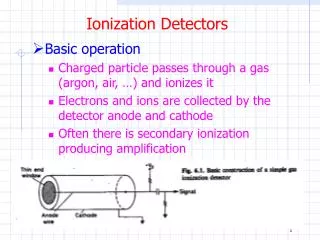
Ionization Chamber • Ionization chambers are a fundamental type of dosimeter in radiation physics • Measurement of the current or charge or reduction in charge gives the exposure or absorbed dose • Free-air ionization chamber • Thimble chamber • Plane parallel chamber • Pocket dosimeter
Ionization Chamber • Current mode • Current gives average rate of ion formation of many particles • Pulse mode • Voltage gives measure of individual charged particle ion formation
Ionization Chamber • Free-air chamber
Ionization Chamber • Used as a primary standard in standards laboratories • Used to measure X • Guard wires and guard electrodes produce uniform electric field • E ~ 100-200V/cm between plates • Assumes CPE • Limited to E<3 MeV (if pressurized) because of electron range
Ionization Chamber • Free-air chambers are not so practical however • Instead one uses an ion chamber with a solid, air equivalent wall
Ion Chambers EXRADIN A12 Farmer EXRADIN A3 Spherical Chamber EXRADIN 11 Parallel Plate Chamber EXRADIN A17 Farmer EXRADIN mini thimble EXRADIN A12 thimble
Ionization Chamber • Vendors Capintec Inc. Nuclear Associates VICTOREEN INC
Ionization Chamber • 0.6 cm3 Farmer chamber
Ionization Chamber Cavity Electrode Sleeve
Ionization Chambers • Materials used • A150 = Tissue equivalent plastic • C552 = Air equivlaent plastic • PMMA = Polymethyl-methacrylate (lucite)
Ionization Chamber • Farmer chamber • Farmer type has a graphite wall and aluminum electrode • For CPE , amount of carbon coating and size of aluminum electrode is adjusted so that the energy response of the chamber is nearly that of photons in free air over a wide range of energies • Since an exact air equivalent chamber and knowledge of V is difficult, in practice they must be calibrated against free air chambers for low energy x-rays • Nominal energy range is 60 keV – 50 MeV
Ionization Chamber • Correction factors • Saturation • Recombination • Stem effects • Polarity effects • Environmental conditions
Ionization Chamber • Need to ensure chamber is used in the saturation region
Ionization Chamber • Stem irradiation can cause ionization measured by the chamber so a correction factor will be needed • Found by irradiating the chamber with different stem lengths in the radiation field
Ionization Chamber • The collection efficiency can be measured by making measurements at two different voltages (one low and one nominal) • Polarity effects can be measured by making measurements at both polarities and taking the average • Environmental conditions are corrected to STP by