Download
1 / 31
330 likes | 653 Views
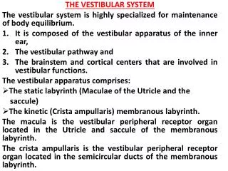
Vestibular System. Vestibular receptors provide information on head orientation and motion, and it’s been highly evolutionarily conserved. . The receptors are in the labyrinth of each inner ear, and they send afferents to secondary neurons in the vestibular nuclei of the brainstem.

E N D
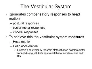
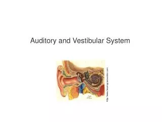
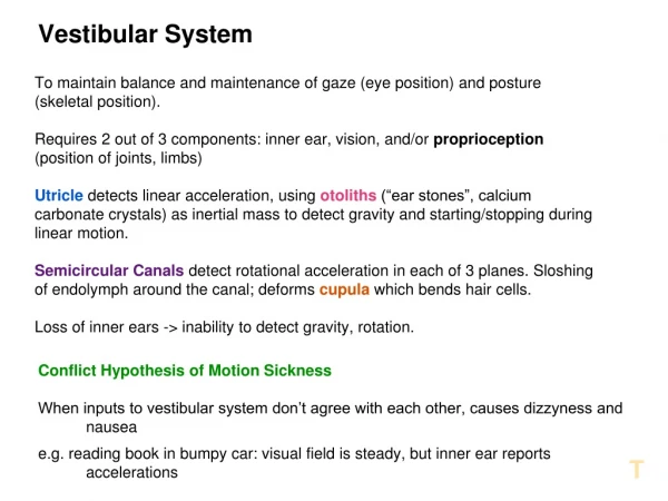
Vestibular receptors provide information on head orientation and motion, and it’s been highly evolutionarily conserved.
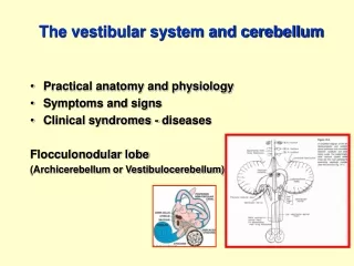
The receptors are in the labyrinth of each inner ear, and they send afferents to secondary neurons in the vestibular nuclei of the brainstem. These project to centers controlling eye, neck and trunk movements.
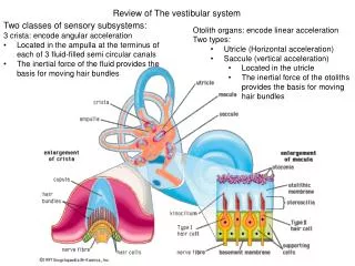
Vestibular receptors include 3 semicircular canals that detect angular acceleration and 2 otolith organs (utricle and saccule) that respond to linear acceleration and orientation.
The vestibular nerve, which gets input from these receptors, has a superior subdivision (utricle, horizontal and superior semicircular canals) and inferior division (saccule and posterior semicircular canals). Each subdivision has a ganglia of Scarpa.
• Head movements control the vestibulocollic (neck, VCR) and vestibulo-ocular (eyes, VOR) reflexes, which stabilize the head and eyes during movement. VOR maintains an image on the fovea during head movement. It is very accurate in normal function.
There is a membranous canal within a bony canal. The membranous canal is filled with endolymph (high K, low Na), and the space between the bone and membrane is filled with perilymph (low K, high Na). Each canal has an ampulla with sensory hair cells, and within the ampulla is a cupula with ciliated receptors.
With head movement, inertia-driven motion of the endolymph in the semicircular canals causes deflection of the cupula, which moves the stereocilia. If they move toward the kinocilium, hair cells release transmitter and discharge afferent nerve fibers.
If they move away from the kinocilium, they release less transmitter and nerves fire less. Afferent nerves have a resting discharge rate, which may be increased or decreased.
L and R horizontal canals are in the same plane. L anterior and R posterior semicircular canals are in the same plane, as are L posterior and R anterior. Movement that leads to excitation in one of these co-planar pairs will lead to inhibition in the other.
The basic vestibulo-ocular reflex is a three-neuron arc. The sensory afferents go from the inner ear to the vestibular nuclei in the brainstem. They synapse with second order neurons that project to extraocular muscle motor nuclei (abducens, trochlear, occulomotor).
These second order neurons go to some motor nuclei ipsilaterally and some contralaterally to coordinate the reflex. So if you move the ipsilateral medial rectus (occulomotor nucleus innervated via tract of Deiters), you will also want that neuron to innervate the contralateral lateral rectus (abducens nucleus) so that both eyes move in the same direction.
Inhibitory connections decrease activity in the antagonist muscles. VOR is very fast and accurate. • VOR is modifiable, so can be calibrated to specific situations and contexts. When you wear bifocals with miniaturizing portions and magnifying portions, the VOR must respond differently in those regions.
This is regulated by motion of images on the retina (retinal slip), which creates blurring called oscillopsia. Retinal slip is a cue for adaptation of the VOR, and the vestibulocerebellum (flocculus and paraflocculus) plays an important role in carrying out the changes.
• Ewald’s laws: Semicircular canals are sense eye and head movements in the same plane as the canal. Stimuli that result in excitation of receptors or afferents are responsible for producing the larger eye and head movements.
• Horizontal canals move the eyes medial/lateral, whereas superior and posterior canals move the eyes up/down with an additional rotation around the visual axis.
• Each otolith organ (utricle/saccule) has a region with sensory epithelium called the macula. The macula’s hair cells have cilia that project into a gelatinous mass with CaCO3 crystals called otoconia.
With linear acceleration, inertia leads to shearing force on the stereocilia. As with the semicircular canals, these cilia moving toward the kinocilium produces an excitatory output, and movement away from the kinocilium produces an inhibitory output.
Whereas hair cells in the ampulla of each semicircular canal are oriented in the same direction, some hair cells in each otolith organ are oriented in opposite directions from one another. So, with a given linear acceleration, some hair cells are excited and others inhibited.
Vertigo is the illusion of motion when none is present, usually a result of vestibular disorder. Oscillopsiais a visualized motion of objects known to be stationary, and if this is induced by head movement it is often indicative of vestibular dysfunction. If VOR doesn’t compensate for head movement, the images move on the retina resulting in oscillopsia.
Nystagmus is a rapid beating of the eyes with slow and fast components. The slow component results from imbalance in the vestibular signals from the two labyrinths. Since movement inhibits semicircular canals on one side and inhibits them on the other, any imbalance in signaling will be interpreted as movement, even if the head is still.
This “movement” will evoke a VOR. The eye will drift slowly toward the side with decreased signaling, then quickly reset. If, as in this case, it occurs with no head movement or external stimulus, it’s termed spontaneous nystagmus.
• Three cardinal features of spontaneous nystagmus: It is horizontal/torsional with fast components moving toward the labyrinth with greater activity. It is more rapid with the patient looks in the direction of the fast components, and diminished if he/she looks in the other direction. It can be suppressed by visual fixation.
Nystagmus can be evoked with a caloric test, in which warm or cold water placed in the external ear lead to convective flow of the endolymph in the semicircular canal.
With warm water, endolymph flows toward the ampulla and results in excitation. Cold water does the opposite. The imbalance in vestibular signaling lead to nystagmus.
Disorders of the labyrinth: o Benign paroxysmal positional vertigo: most common cause of vertigo. Otoconiaare dislodged and pulled most often into the posterior semicircular canal. Motion of these particles causes nystagmus in the plane of the affected canal (usually vertical/torsional).
Slow components are down with superior poles moving away from the affected ear. This can be cured with maneuvers to reposition the particles.
Vestibular neuritis: sudden onset vertigo that lasts for days. It usually affects the superior division of the vestibular nerve, affecting the horizontal and superior canals and utricle.
Meniere’s syndrome: fluctuating sensorineural hearing loss, tinnitus (ringing), fullness of ears, and episodic vertigo. Due to distention of endolymphatic spaces with repture of the labyrinthine membranes. Controlled with low salt diet and diuretics.
Superior canal dehiscence syndrome: caused by an opening on the bone over the semicircular canal. Vertigo and oscillopsia induced by noise or changes in middle ear or cranial pressure. These cause movements in the plane of the affected canal (usually superior canal).
o Bilateral vestibular hypofunction: often caused by gentamicintoxicity. Oscillopsiaoccurs. Vestibular hair cells take up gentamicin, which chelatesiron. The complex with iron forms radicals that are toxic to hair cells.