Download
1 / 22
240 likes | 306 Views
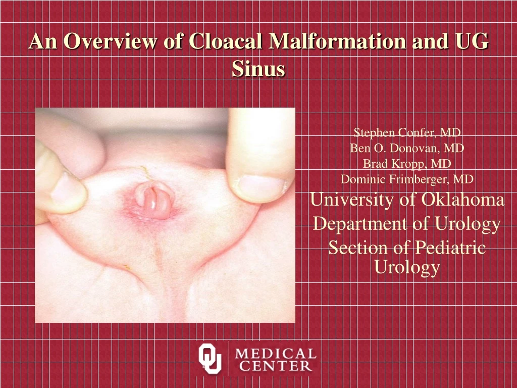
An Overview of Cloacal Malformation and UG Sinus. Stephen Confer, MD Ben O. Donovan, MD Brad Kropp, MD Dominic Frimberger, MD University of Oklahoma Department of Urology Section of Pediatric Urology. Normal GU Development.

E N D
An Overview of Cloacal Malformation and UG Sinus Stephen Confer, MD Ben O. Donovan, MD Brad Kropp, MD Dominic Frimberger, MD University of Oklahoma Department of Urology Section of Pediatric Urology
Normal GU Development The mesonephric (Wolffian) duct descends from the mesonephros to meet the urogenital sinus. Once this connection is made, fetal urine drains into the urogenital sinus. The ureteric bud arises from the mesonephric duct and progresses laterally to invade the metanephrogenic blastema (precursor of the mature kidney). The caudal end of the mesonephric duct (past the ureteric bud is called the common excretory duct. As renal development proceeds, the common excretory duct is incorporated into the urogenital sinus.
Normal GU Development Progressive incorporation of the common excretory duct eventually leads to separate openings of the ureter and mesonephric duct into the urogenital sinus. By 37 days of gestation, the ureter empties into the urogenital sinus cephalad to the mesonephric duct. The urogenital sinus is divided between the orifices of these two tubes. The cephalad portion of the urogential sinus will become the bladder while the caudal portion will become the urethra
UG Sinus • Persistent UG Sinus with normal anus and rectum 2° to adrenogenital syndrome • Genetic female with varying degrees of masculinization of lower GU tract 2° excess androgens • Least severe: clitoral hypertrophy and labio-scrotal fusion • Severe phallus: resemble penis and a high vagina that enters close to bladder neck
SEVERITY less more UG Sinus Normal appearing vulva in an infant Retraction of labia reveals only a single opening Karyotype 46 XX. 12 years old girl. Penis-like clitoris.
Exam • Inspect perineum • anal opening in normal to anterior position • Single opening anterior to rectum • Clitoromegaly consider adrenogenital syndrome • External Genitalia masculinized • penis & scrotum • no palpable gonads • Important: newborn w/o palpable gonads should increase suspicion that the patient could be female • Rectal examination may reveal a uterus
Work Up • Exam • Labs • U/S • Genitogram • Cystosccopy
Surgical Correction • Determine level where urethra and vagina converge • Classical Mobilize vagina and urethra separately • Mobilize entire UG sinus (Pena)
Low Confluence • Mobilize entire UG sinus less devascularization of vagina • Vaginoplasty- with posterior perineum flaps • Urethroplasty +/- use UG sinus create neobladder, gives appearance of vagina with urethra • Reconstruction of External Genitalia • Labia minora UG sinus • Labia majora • Clitoral reduction
High Confluence • Vagina enters above level of external sphincter • Vagina separated from urethra • Vaginoplasty- mobilize vagina and create perineum flaps • Urethroplasty use UG Sinus for distal urethra
Cloacal Anomalies • 7 mm embryo cloaca common chamber where GI and urinary tract converge • Latin mean sewer • 22mm urorectal septum divides cloaca into UG sinus and rectum • Persistent cloaca normal in birds and reptiles • Interruption of differentiation give a wide range of abnormalities
Cloacal Malformation • 1:50,00 births • Bladder, vagina, and rectum converge above the perineum • When convergence is low can have a normal looking female with anteriorly placed or imperforate anus • Conservation of structures at the bladder neck • Urine can fill vagina and displace bladder anteriorly causing obstruction and hydronephrosis • Obstruction of colon
Cloacal Malformation • Cloaca exists when the rectum, vagina and urinary tract meet and fuse into a single common channel. • If the channel is short (<3cm), well-developed sacrum and good sphincters will likely be present. • A longer channel indicates a more complex defect with a poor sphincter mechanism and an abnormal sacrum
Cloacal Malformation • Preoperative photos show a normal appearing external genitalia, but no anus. Examination of the vaginal introitus reveals a rectal fistula to the posterior vaginal fourchette through which a catheter was placed for demonstration
Prenatal Diagnosis • A fetal ultrasonogram at 27 weeks' gestation; the most obvious feature is the large cystic structure arising from the fetal pelvis. This is a dilated vagina (hydrocolpos) which is compressing the bladder inferiorly.
Prenatal Diagnosis • A maternal MRI scan with intravenous contrast medium at 31 weeks' gestation. The larger cystic structure seen in the fetal pelvis is a dilated vagina (hydrocolpos) filled with fetal urine; the other cystic structure situated to the right of the view is the bladder
Other Findings • Sacral bony abnormalities seen on plain films
Management • Decompression with right transverse colostomy • Resist pull through • Catheterize probably vagina, seldom vesicostomy or vaginotomy • Treat co-morbid condition • W/U: electrolytes, U/S, genitogram, cystoscopy MRI (R/O tethered cord) • Treat urinary tract abnormality first or at the same time
Management • VUR 84/135 • Colostomy in neonate to releive colon obstruction • CIC, seldom vesicostomy or vaginotomy • Definitive repair until 1 y/o • Pt with double vaginas can have septum incised endoscopically
Surgical Repair • Position- total body prep alternating position • Posterior sagittal approach • Mobilze rectum and pull through • Mobilize vagina and separate from UG-sinus or bladder neck, rotate 90° prevent fistulas, may need perineum flaps or bowel interposition • Mobilize UG Sinus to create neourethra • Post need CIC, enemas • Colostomy closure 6 weeks
Hendron • 154 pts • Voiding • 83 spontaneous • 40 CIC , 1 continent diverson, 4 UD, 5 wet, 8 too early • Bowel • 83 move bowels spontaneously • 38 enema, 9 colostomy, 7 soil, 5 too early • Coitus 24 adults, • 14 married, 7 no coitus ( 2 married) • 6 conception caesarian section