Download
1 / 19
190 likes | 206 Views
Trauma. Mohammad Jomaa. Introduction. Forms of injury include: Foreign bodies becoming lodged under the upper lid or on the surface of the eye, especially the cornea.

E N D
Trauma Mohammad Jomaa
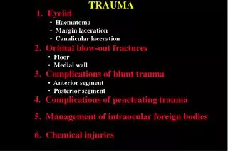
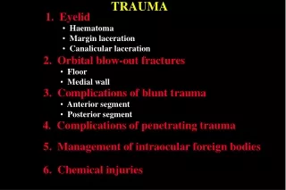
Introduction Forms of injury include: • Foreign bodies becoming lodged under the upper lid or on the surface of the eye, especially the cornea. • Blunt trauma from objects small enough not to impact on the orbital rim (shuttlecocks, squash balls, champagne corks and knuckles are some of the offenders). The sudden alteration of pressure and distortion of the eye may cause severe damage. • Penetrating trauma, where ocular structures are damaged by a foreign body which passes through the ocular coat and may be retained in the eye. With the introduction of the seat belt laws the incidence of penetrating injury following road traffic accidents has declined. • Chemical and radiation injury, where the resultant reaction of the ocular tissues causes the damage.
History, symptoms and signs A careful history is essential. • Use of a hammer and chisel can release a flake of metal which will penetrate the globe, leaving only a tell-tale subconjunctival haemorrhage to indicate penetration of the sclera and suggest a retained foreign body. Pain may be minor and heat, generated by the high velocity, sterilizes the fragment so that infection does not occur. Undetected, such a retained foreign body may have devastating effects on the eye. • A wire under tension, or a rose thorn, may penetrate the cornea briefly, sometimes creating a barely visible track. • A blunt injury to the eye may also result in damage to the orbit (blow–out fracture). • It is vitally important to determine the nature of any chemical that may have been in contact with the eye. Strong alkalis and acids penetrate the anterior tissues of the eye and may rapidly cause irreversible damage. • As in all history taking, it is essential to enquire about previous ocular andmedicalevents
Examination • Without a slit lamp • The examination will depend on the type of injury. In all cases it is important that visual acuity is recorded in the injured and uninjured eye for medico – legal reasons, among others. • Where a penetrating injury is suspected and pressure to the globe must be avoided, it may only be possible to measure vision approximately in the injured eye; the patient may be able to detect light shone through the closed lid and even the direction of the source. • The skin around the orbit and eyelids should be carefully examined for a penetrating wound.
Orbital injury • Damage to the orbit itself (a blow-out fracture) is suspected if the following signs are present: • Emphysema (air in the skin which crackles when pressed) derived from a fractured sinus. • A patch of paraesthesia below the orbital rim suggesting infraorbitalnerve damage. The infraorbital nerve is commonly injured in orbital blow – out injury involving the floor of the orbit. • Limitation of eye movements, particularly on upgaze and downgaze, due to trapping of the inferior rectus muscle by connective tissue septa caught in the fracture site in the inferior orbital floor, the wall most commonly fractured. • Subsequently the eye may become recessed into the orbit (enophthalmos). • If the lid margin is cut at the medial canthus it is important to determine if either of the lacrimal canaliculi is severed. This will cause epiphoraif untreated.
Further examination of a traumatized eye will require the instillation of a local anaesthetic to facilitate lid opening (lidocaine, amethocaine). If a penetrating eye injury is suspected it is important that no pressure is applied to the globe, to avoid expression of its contents.
The conjunctiva and sclera • These must be examined for the presence of any lacerations. If the history is appropriate a subconjunctival haemorrhage should be considered to be the possible site of a scleral perforation. The fundus should be examined with full mydriasis to exclude a retained intraocular foreign body. • Retained, iron-containing foreign bodies may have an insidious and particularly devastating effect on the eye. A tiny, high-velocity metal fragment, penetrating the ocular coats and retained in the peripheral vitreous cavity can, with time, lead to a progressive, pigmentary degeneration of the retina. A discolouration of the iris (heterochromia) , a fixed mydriasis and cataract can be a late clues to the diagnosis. Failure to detect and remove such a foreign body at the time of injury results in irreversible blindness. The mechanism of damage is by the diffusion of ferrous ions throughout the globe and the generation of free radicals, via the Fenton reaction, in affected tissues. • Events of this kind emphasize the need to wear protective goggles when using metal hammers or hammering on metal.
If a chemical injury has occurred, the conjunctiva may appear white and Ischaemic. If such changes are extensive, involving the greater part of the limbal circumference, corneal healing will be grossly impaired because of damage to the epithelial stem cells of the cornea, which are located at the limbus. There will be additional complications such as uveitis, secondary glaucoma and cataract.
The cornea • This is examined for loss of the epithelial layer (abrasion), for lacerations and for foreign bodies. The instillation of fluorescein will identify the extent of an abrasion and use of concentrated fluorescein, will identify a leak of aqueous through a penetrating wound. If the globe appears intact and asubtarsal foreign body is suspected (signalledby fi ne, staining, vertical, linear corneal abrasions) the upper lid must be everted, to expose the underside of the Lid. This allows any foreign body to be identified and removed. • Electromagnetic radiation may injure the conjunctiva and the cornea. Unprotected exposure to ultraviolet radiation from an arc lamp (arc eye) or sunlamp, or reflected from snow, is the commonest cause of this severely painful condition. Typically, severe ocular pain onsets acutely, 6 hours after exposure to the radiation, and the cornea shows diffuse epithelial oedema and punctate erosions. These resolve within 24 – 48 hours.
The anterior chamber • Blunt trauma may cause haemorrhage into the anterior chamber, where it collects with a fluid level, visible as a hyphaema. This is caused by rupture of the root of the iris blood vessels, or the iris may be torn away from its insertion into the ciliary body (iris dialysis) to produce a D-shaped pupil. Hyphaema may also be seen with a penetrating eye injury and the shape of the pupil may be distorted if the peripheral iris has plugged a penetrating corneal wound. The pupil may also show a fixed dilatation as a result of blunt trauma (traumatic mydriasis).
The lens • Dislocation of the lens following blunt trauma may be suggested by a fluttering of the iris diaphragm on eye movement (iridodonesis). Lens clarity should be assessed with the slit lamp and against the red reflex after pupil dilatation. • Cataracts develop abruptly with direct penetrating trauma. • Blunt trauma also causes a posterior subcapsular cataract within hours of injury, which may be transient.
The fundus • The fundus should be inspected with a direct ophthalmoscope after full mydriasis. If no systemic neurological complications accompany the injury and ocular penetration is not suspected, the pupil can be dilated. Areas of retinal haemorrhage may be seen and typical patches of white, retinal oedema (commotio retinae). A retinal dialysis (a separation of the peripheral retina from its junction with the pars plana of the ciliary body) and a macular hole may also result from blunt trauma. The choroid may also become torn, causing a subretinal haemorrhage which later leads to subretinal scarring. Peripheral retinal changes can only be excluded with indirect ophthalmoscopy or slit - lamp microscopy. If there is no red reflex and no fundus details are visible, this suggests a vitreous haemorrhage. • The optic disc may be pale from a traumatic optic neuropathy caused by avulsion of the blood vessels supplying the optic nerve. Although this is uncommon, it leads to a profound loss of vision and no treatment is available.
With a slit lamp • The slit lamp allows a more detailed examination to be performed, which may reveal: • a shallow anterior chamber compared to the fellow eye, suggesting an anterior penetrating injury with aqueous loss; • a microscopic hyphaema, where red cells are present, circulating in the anterior chamber, but have not yet settled to form a level; • presence of white cells in the anterior chamber (traumatic uveitis); • recession of the iridocorneal angle seen with a gonioscopic contact lens (the ciliary muscle apex is disinserted from the scleral spur and moves posteriorly) – this may be seen with blunt trauma and results in raised intraocular pressure, sometimes after a delay of months or years; • raised intraocular pressure measured by applanation tonometry – this may accompany a hyphaema, lens dislocation or, as noted, damage to the chamber angle.
Treatment • Lacerations to the skin and lids These require careful apposition and suturing, particularly if the lid margin is involved, to retain the lid contour. If one of the lacrimal canaliculi is damaged an attempt can be made to repair it • Corneal foreign bodies Corneal foreign bodies should be removed with a needle under topical anaesthesia . Subtarsal objects can often be swept away with a cotton-wool bud from the everted lid. The patient is then treated as for an abrasion. If there is any suggestion that a foreign body may have penetrated the globe the eye must be carefully examined with dilation of the pupil to allow a good view of the lens and retina. A radiograph of the orbits, with the eyes looking up and then down, or a CT scan, may also be indicated if an intraocular foreign body is suspected. Microsurgical techniques can be used to remove foreign bodies from the eye under direct view..
Corneal abrasions This is an extremely painful condition which normally heals rapidly. It should be treated with antibiotic ointment, with or without an eye pad. Dilatation of the pupil with cyclopentolate 1% can help to relieve the pain caused by spasm of the ciliary muscle. In some cases, when such an injury is caused by flexible objects such as Fingernails, twigs or the edge of a newspaper, a minority of patients experience recurrent episodes of pain, particularly in the early hours of the morning or on waking. This condition is termed recurrent corneal erosion and is due to a defective adhesion of the resurfacing epithelium to Bowman ’ s layer at the site of injury. Prophylaxis against further recurrent corneal erosions is attempted, using a lubricating ointment at night for several weeks after an initial attack, but more permanent results are achieved by inducing a subepithelial scar at the site of the original injury to reinforce the attachment of the basal epithelial layer to Bowman’s layer. This can be induced by laser treatment or by applying a series of micropunctures with a needlepoint to the affected zone. UV injury to the cornea responds quickly to topical steroids.
Corneal and scleral penetrating trauma Once identified, no further examination of the globe should be performed but a shield should be gently placed over the eye and the patient referred for urgent ophthalmic treatment. These serious injuries, often with grave implications for sight, require careful microsurgical suturing to restore the integrity of the globe. Once the eye has settled from this primary repair additional operations are often required later, to: • remove a foreign body; • remove a cataract; • replace a corneal opacity with a corneal graft; • repair a detached retina or remove the vitreous gel to prevent detachment. • Uveitis This responds to the usual treatment with steroids and dilating drops. It may be accompanied by elevated intraocular pressure requiring additional medical treatment.
Hyphaema This usually settles with rest, but a rebleed may occur in the first 5 – 6 days after injury. Steroid eye drops are given for a short time, together with dilation of the pupil. Steroids reduce the risk of rebleeds. The commonest complication is a raised ocular pressure (It is for this reason that rest is important). Raised pressure is due to the accumulation of empty, red cell ‘ ghosts ’ in the trabecular meshwork or to damage to the drainage angle itself It usually responds to medical treatment, but occasionally surgical intervention is required
Retinal damage • In commotio retinae the affected zone of retina opacifies and obscures the underlying choroidal detail. It usually resolves, but requires careful observation since retinal holes may develop in affected areas and may lead to subsequent retinal detachment. • Retinal dialysis requires surgical intervention to repair any detached retina. • A vitreous haemorrhage may absorb over several weeks, or may require removal by vitrectomy. • Chemical injury • The most important part of the treatment is to irrigate the eye immediately with copious quantities of clean water at the time of the accident. This must be repeated when ophthalmic care is available, when it is also important to irrigate under the upper and lower lid to remove solid particles, e.g. lime. The nature of the chemical can then be ascertained by history and measuring tear pH with litmus paper. Administration of steroid and dilating drops may be required. Vitamin C, given both orally and topically, may improve healing. Systemic and topical anticollagenases may be needed (e.g. tetracyclines). • Extensive damage to the limbus may prevent resurfacing of the cornea with epithelium. A prolonged epithelial defect may lead to a corneal ‘ melt ’ (keratolysis). This is treated later, by limbal stem cell transplantation , for instance from the normal, fellow eye or from a donor source or oral mucosal cells.
Orbital blow-out fracture If a blow - out fracture is suspected, a CT scan will delineate the bony and soft - tissue injury. If this is not possible then plain orbital radiographs are performed. Treatment may be delayed until the periorbital swelling has settled. At this later stage the degree of enophthalmos and the limitation of eye movementcan be measured. Although some surgeons advocate an early intervention to obtain the best results, many patients will require no surgery at all.