Download
1 / 33
330 likes | 355 Views
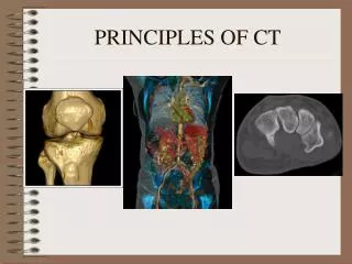
CT PRINCIPLES. AYMAN OSAMA. Starting with 1st and 2nd generations that combined rotational and translatory pencil beam and partial fan beam scanning followed by 3rd and 4th generation full continuous rotation around 360°.

E N D
CT PRINCIPLES AYMAN OSAMA
Starting with 1st and 2nd generations that combined rotational and translatory pencil beam and partial fan beam scanning followed by 3rd and 4th generation full continuous rotation around 360°
spiral CT has dramatically changed the performance of body CT scans.The elimination of respiratory misregestration artifacts, the minimization of motion artifacts and the production of overlapping images without additional radiation exposure are the most important technical advantages of helical CT.
The technique involves the continuous acquisition of projection data through a 3D volume of tissue by continuous rotation of the x-ray tube and detectors and simultaneous translation of the patient through the gantry opening
Nearly one decade after helical CT introduction, another technical breakthrough has occurred that promises to be equally important. Multi-detector row CT (MDCT) offers better longitudinal and temporal resolution that benefits nearly all imaging applications
In 1992, dual-section helical scanner was created, the first and simplest multisection scanner.
In the fall of 1998, several equipment manufacturers launched the next generation of multisection CTscanners.
These units have four data acquisition systems connected to multi-detector arrays to provide a "quad-section" CT scan, increasing the speed of data collection by a factor of four and gantry rotation speed by a factor of two over conventional single-section helical CT scanners.
In simple terms, multi-detector row CT (also called multi-slice CT, multichannel CT, or multisection CT) allows acquisition of multiple slices with a single gantry rotation. MultidetectorCT’, is somewhat misleading, as the number of detector rows is generally larger than thenumber N of slices. The latter, however, is the decisive feature of such a scanner.
MultidetectorCT’, is somewhat misleading, as the number of detector rows is generally larger than thenumber N of slices. The latter, however, is the decisive feature of such a scanner
The conventional single-slice helical CT scanner has one x-ray tube and a single row of detectors. This detector row contains 500–900 detector elements, which describe an arc in the transverse (axial or x-y) plane, providing one channel of spatial data
The multi-detector row CT scanner has one x-ray tube and multiple rows of detectors along the longitudinal (z) axis of the patient.
Each row has 500–900 elements, and many rows together create a two-dimensional curved array containing thousands of detector elements, which are connected to four up to thirty two data acquisition systems that generate four to thirty two CT slices in each gantry rotation
To register four slices simultaneously, a minimum of four detectors must be placed side by side along the Z-axis, but theoretically this will give us a machine that gives four slices of fixed section thickness that can not be changed. So, to offer a choice of several section thicknesses, more than four detector elements along the Z-axis are required.
(1) Matrix detectors: where all detectors are of the same size. There is 16 identical detectors arranged side by side in the Z-axis, each one of them is 1.25 mm making it covering a distance of 20 mm. (General Electric)
(2) Adaptivearray detectors: where they vary from thinner inside to thicker outside. There is 8 detectors, varying in size from 1 mm to 5 mm. They have a mirrior image arrangement with the 1mm detector in the middle, then the 1.5 mm detector, then the 2.5 mm detector, and finally the 5 mm detector. These detectors arrangement cover also 20 mm in the Z-axis direction. (Siemens and Philips)
(3) Hybrid detectors: where usually two sizes are used with the thinner detectors located centrally. There are 34 detectors, 30 of them are 1 mm in length and the other 4 detectors are 0.5 mm in length. They are arranged with the smaller 4 detectors in the middle and other 30 on both sides, covering a distance of 32mm in the Z-axis direction. (Toshiba)
Multi-detector row CT scanners alter the acquisition thickness by varying the number of detectors used. For example, a four-slice scanner using the matrix design has a Z-axis width of 20 mm subdivided into 16 equal parts of 1.25 mm each
Scanning Speed • Single-section helical CT scanners have generally had a 360° gantry rotation speed of 1 second. With multisection CT, some scanners offer a gantry rotation speed of 0.5 second, twice as fast as that of single-section helical CT. Because multisection CT scanners can generate up to four sections per revolution, they are up to eight times faster than single-section helical CT scanners.
To scan the same volume in the same time with single-section helical CT, one must increase the pitch or section thickness , thereby degrading image quality.