Download
1 / 35
380 likes | 500 Views
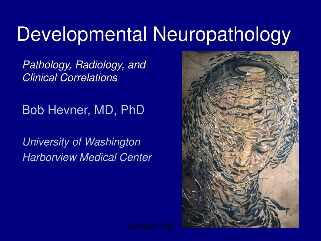
Developmental Neuropathology. Pathology, Radiology, and Clinical Correlations Bob Hevner, MD, PhD University of Washington Harborview Medical Center. Salvador Dali. Classification & Overview. Malformations

E N D
Developmental Neuropathology Pathology, Radiology, andClinical Correlations Bob Hevner, MD, PhD University of Washington Harborview Medical Center Salvador Dali
Classification & Overview • Malformations • Neural tube defects: anencephaly, craniorachischisis totalis, encephalocele, spina bifida (myelomeningocele) • Patterning defects (holoprosencephaly) • Cell migration disorders: lissencephaly, polymicrogyria, heterotopia • Gyral abnormalities: Down syndrome, thanatophoric dysplasia • Cerebellar anomalies: Chiari malformations, syrinx • Spinal cord lesions: diastematomyelia, hydromyelia, spinal lipoma • Hypoxic/ischemic lesions • White matter: periventricular leukomalacia (PVL) • Gray matter: ulegyria, status marmoratus • White and gray: schizencephaly, multicystic encephalopathy • Hemorrhages • Mid-gestational: germinal matrix hemorrhage • Perinatal: cerebellar, choroid plexus, venous thrombosis • Metabolic disorders • Leigh’s disease • Autism
Normal fetal brain development 17 wk 22 wk 28 wk • Morphogenesis (gyration) • Growth • Cell migrations • Axon connections • Myelination 30 wk
Quantitative Measurements • Brain weight (overall growth) • Cerebral mantle thickness (ventriculomegaly)
Neural tube defects • Failure of cranial or spinal patterning, closure, or differentiation • Neural tube closure starts at three different points • Potential for a great variety of defects due to failure of this process • Teratogens (valproic acid) • Genetic factors • Prevention: folic acid (19-70% reduction)
Anencephaly • “Frog-like” appearance • Begins as exencephaly, with cranial defect; neural tissue destroyed by exposure to amniotic fluid. • Remaining tissue: “area cerebrovasculosa”
Craniorachischisis totalis Complete failure of cranial and spinal neural tube or mesodermal (bony) closure. Early embryonic defect.
Encephalocele Herniation of brain tissue through cranial defect. Occipital is most common in Western countries. Frontal is more common in Southeast Asia.
Spinal Dysraphism A: Open myelomeningocele B: Closed myelomeningocele C: Dermal sinus D: Sacral agenesis (often hydromyelia, skin dimple)
Myelomeningocele Open or barely closed neural tube defect. Most often occur in lumbosacral region. “Areamedullovasculosa” Associated withChiari type II
Holoprosencephaly • Defect of dorsovental patterning in the embryonic forebrain. • Frequently associated with facial anomalies (cyclopia, proboscis) • Associated with mutations of Shh, Zic2, other genes • Lobar • Semilobar • Alobar • Lobar • Semilobar • Alobar
Holoprosencephaly • Often diagnosed prenatally (23 wk GA) • Lobar • Semilobar • Alobar Anterior Posterior
Neuronal Migration Disorders • Lissencephaly • “Smooth brain” with reduced number/absence of gyri and sulci • Synonyms: agyria (absence of gyri), pachygyria (thick gyri) • Polymicrogyria • Increased number of tiny gyri with microscopic fusion of layer 1 • Must be distinguished from increased gyral complexity, e.g., in hydrocephalus • Heterotopia • Laminar • Periventricular
Migration Disorders:Polymicrogyria Diverse etiologies (genetic, hypoxic/ischemic, metabolic) Frequently associated with epilepsy
Migration Disorders: Lissencephaly aka agyria/pachygyria Type I: smooth surface -Miller-Dieker (17p-), LIS1 Type II: irregular surface with glioneuronal heterotopias (subarachnoid)
Gyral Anomalies: Thanatophoric Dysplasia Lateral view • Affected individuals also have severe dwarfism. • Excessive temporal lobe growth. “Bear claw”. • Caused by mutation of FGFR3, which regulates growth of bones and brain. Inferior view
Chiari malformations • Type I • Cerebellar tonsils extend through foramen magnum into cervical spinal canal (chronic tonsillar herniation) • 40-75% associated with cervical cord syringomyelia • Usually becomes symptomatic in teen to early adult years • Type II • Arnold-Chiari malformation • Cerebellar vermis and hemispheres extend through foramen magnum into cervical spinal canal • Associated anomalies of brainstem (100%) • Associated myelomeningocele (95%) • Type III • Cerebellar encephalocele • Very rare • Type IV • Cerebellar aplasia/absence • May be secondary destructive lesion
Chiari type II (Arnold-Chiari) • Pathogenesis unknown. • Cerebellar vermis herniation associated with lumbar myelomeningocele. • “Beaking” of the tectum • Flattened skull base • Kinking of the medulla • Hydrocephalus 22 wk fetus
Spinal anomalies: Diastematomyelia Partial duplicationof the spinal cord. Defect of spinal cord patterning.
Hydromyelia Dilatation of thecentral canal. Often associated with sacral agenesis. Asymptomatic. Distinguished from syrinx by presenceof ependyma
Spinal Lipoma Sometimes associated with dysraphism. Reflects multipotentiality of “tail bud” embryonic tissue (cf. sacrococcygeal teratoma)
Acquired developmental lesions: susceptibility Susceptibility to hypoxia-ischemia,hemorrhage, etc. depends on age
Hypoxic-ischemic lesions • White matter • Periventricular leukomalacia (PVL): essentially, white matter infarcts • Gray matter • Ulegyria: mushroom gyri, atrophic at depths of sulci • Status marmoratus: “marbled state” of thalamus and basal ganglia, due to hypoxic injury with aberrant myelination • Gray and white matter: large destructive lesions • Schizencephaly and porencephaly • Multicystic encephalopathy
Periventricular leukomalacia: histology • Peak incidence 24-32 weeks gestation • Intrauterine hypoxia/ischemia • Basically a white matter “watershed” infarct • Fetal white matter is particularly susceptible
Ulegyria • “Mushroom” gyri • Actually a response to perinatal hypoxia/ischemia • In swollen brain, cortex at depths of sulci loses blood flow
Germinal matrix hemorrhage • Peak incidence 28-33 gestational weeks • Associated with premature birth • Can cause death when severe • May resolve with no sequelae
Germinal matrix hemorrhage: Grading • Grade 1 • Isolated subependymal hemorrhage (SEH) • Grade 2 • SEH with intraventricular hemorrhage (IVH) but no ventricular enlargement • Grade 3 • SHE + IVH + enlarged ventricles • Grade 4 • SHE + IVH + extension of hemorrhage into brain tissue
Pontosubicular necrosis • Peak incidence around term • Perinatal hypoxia/ischemia • Neuronal death • Morphologically identical to programmed cell death (i.e. ) apoptosis
Ferrugination Neuronal • Neuronal cell death • Typical response of single neurons in infants
The most common malformation? Microcephaly