Download
1 / 39
471 likes | 2k Views
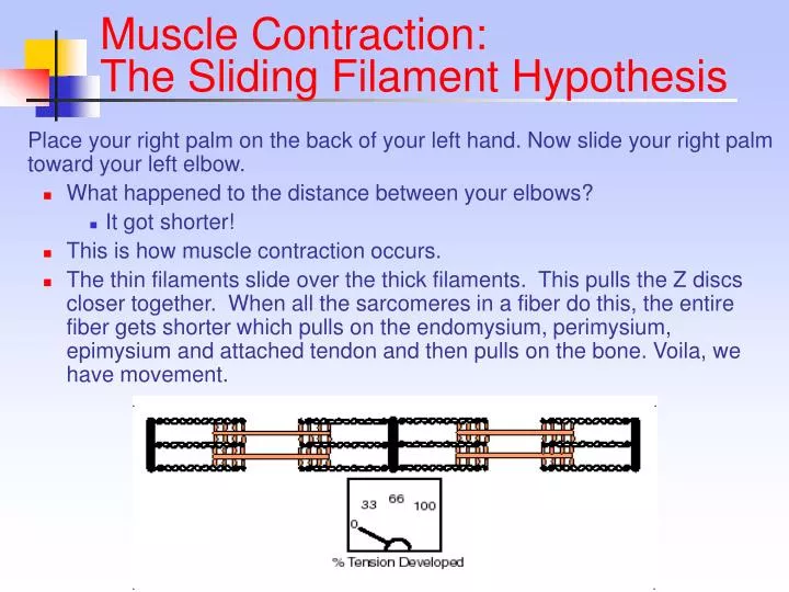
Muscle Contraction: The Sliding Filament Hypothesis Place your right palm on the back of your left hand. Now slide your right palm toward your left elbow. What happened to the distance between your elbows? It got shorter! This is how muscle contraction occurs.

E N D
Muscle Contraction: The Sliding Filament Hypothesis Place your right palm on the back of your left hand. Now slide your right palm toward your left elbow. • What happened to the distance between your elbows? • It got shorter! • This is how muscle contraction occurs. • The thin filaments slide over the thick filaments. This pulls the Z discs closer together. When all the sarcomeres in a fiber do this, the entire fiber gets shorter which pulls on the endomysium, perimysium, epimysium and attached tendon and then pulls on the bone. Voila, we have movement.
Muscle Contraction: The Sliding Filament Hypothesis Here are 2 electron micrographs of the same sarcomere. Do you see the Z discs, A band, H zone, M line, and I bands? How do the 2 pictures differ? What happened?
All the sarcomeres in a fiber will contract together. This contracts the fiber itself. The number of fibers contracting will determine the force of the contraction of the whole muscle. We can actually divide the whole process of muscle contraction into 4 steps: Excitation Excitation-contraction coupling Contraction Relaxation Sliding Filaments
All cells have a voltage difference across their plasma membrane. This is the result of several things: The ECF is very high in Na+ while the ICF is very high in K+. The PM is impermeable to Na+ but slightly permeable to K+. As a result, K+ is constantly leaking out of the cell. In other words, positive charge is constantly leaking out of the cell. Excitation
Excitation • In general each muscle is served by one nerve – a bundle of axons carrying signals from the spinal cord to the muscle. • Within the muscle, each axon will go its own way and eventually branch into multiple small extensions called telodendria. Each telodendrium ends in a bulbous swelling known as the synaptic end bulb.
Excitation The site of interaction btwn a neuron and any other cell is known as a synapse. The synapse btwn a neuron and a muscle is known as the neuromuscular junction.
The minute space between the synaptic end bulb and the sarcolemma is known as the synaptic cleft. There is a depression in the sarcolemma at the synaptic cleft known as the motor end plate. Excitation The synaptic end bulb is filled with vesicles that contain the neurotransmitter, acetylcholine. The motor end plate is chock full of acetylcholine receptors.
The cycle of attachment, power stroke, and release continues as long as calcium and ATP remain available. Typically half the myosin molecules at any time are bound to the actin while the other half are preparing to bind again. A common analogy is climbing a rope hand over hand. Contraction
Is a function of: The number of crossbridges that can be made per myofibril The number of myofibrils per muscle fiber The number of contracting muscle fibers Contraction Strength
Muscle Metabolism • The chemical energy released by the hydrolysis of ATP is necessary for both muscle contraction and muscle relaxation. • Muscles typically store limited amounts of ATP – enough to power 4-6sec of activity. • So resting muscles must have energy stored in other ways.
ATP is necessary for cellular housekeeping duties. ATP powers the combination of glucose monomers (which have been taken up from the blood stream) into the storage polymer glycogen. ATP is used to create another energy storage compound called creatine phosphate or phosphocreatine: ATP + Creatine ADP + Creatine-Phosphate this reaction is catalyzed by the enzyme creatine kinase ATP Use in the Resting Muscle Cell
As we begin to exercise, we almost immediately use our stored ATP. For the next 15 seconds or so, we turn to the phosphagen system, a.k.a., the energy stored in creatine-phosphate. Creatine-P + ADP Creatine Kinase Creatine + ATP The ATP is then available to power contraction and relaxation: myosin ATPase, Ca2+ ATPase in the SR membrane, and Na+/K+ ATPase in the sarcolemma. The phosphagen system dominates in events such as the 100m dash or lifting weights. Working Muscle
After the phosphagen system is depleted, the muscles must find another ATP source. The process of anaerobic metabolism can maintain ATP supply for about 45-60s. Anaerobic means “without air,” and it is the breakdown of glucose without the presence of oxygen. It usually takes a little time for the respiratory and cardiovascular systems to catch up with the muscles and supply O2 for aerobic metabolism. Working Muscle
Anaerobic Metabolism • Lactic acid typically diffuses out of muscles into the blood stream and is taken to the liver, kidneys, or heart which can use it as an energy source. • Anaerobic metabolism is inefficient. Large amounts of glucose are used for very small ATP returns. Plus, lactic acid is a toxic end product whose presence contributes to muscle fatigue. • Anaerobic metabolism dominates in sports that requires bursts of speed and activity, e.g., basketball.
Occurs when the respiratory and cardiovascular systems have “caught up with” the working muscles. Prior to this, some aerobic respiration will occur thanks to the muscle protein, myoglobin, which binds and stores oxygen. During rest and light to moderate exercise, aerobic metabolism contributes 95% of the necessary ATP. Compounds which can be aerobically metabolized include: Pyruvic acid (made via glycolysis), fatty acids, and amino acids. Aerobic Metabolism
Aerobic Metabolism • It occurs in the mitochondria. • Pyruvic acid from glycolysis is the primary substrate. The cell also utilizes fatty acids and amino acids. • Aerobic respiration typically yields 36 ATP per molecule of glucose. Compare this to anaerobic metabolism.
Muscle Fatigue • Physiological inability to contract • Results primarily from a relative deficit of ATP. • Other contributing factors include the decrease in sarcoplasmic pH (what causes this?), increased sarcoplasmic [ADP], and ionic imbalances.
Refers to the fact that post-exercise breathing rate >>> resting breathing rate This excess oxygen intake serves many tasks: Replenish the oxygen stored by myoglobin and hemoglobin Convert remaining lactic acid back into glucose Used for aerobic metabolism to make ATP which is used to: Replenish the phosphagen system Replenish the glycogen stores Power the Na+/K+ pump so as to restore resting ionic conditions within the cell. Oxygen Debt
Motor Units A motor unit is defined as a somatic motor neuron and all the skeletal muscle fibers it innervates. When this neuron is stimulated, all the muscle fibers it synapses upon will be stimulated and will contract as a unit The # of muscle fibers per motor unit may be as high as several hundred or as few as four. • The smaller the motor unit, the finer and more delicate the movements. • Extraocular muscles typically have small motor units while the large postural muscles have large motor units
Motor Units Notice that the muscle fibers of a single unit are not clustered together but are spread out. What’s the advantage to this?
It should be obvious that you can contract a muscle at just about any rate and with any force you desire. How does this fact concur with the quickness of a single muscle twitch. We achieve smooth contractions of the whole muscle by varying the frequency of stimuli sent to the muscle fibers and by recruitment – varying the number and size of the motor units involved Graded Responses Thought problem: compare the act of picking up a pencil with the act of picking up a desk
Types of Contractions Contractions can be: Isometric Iso= same, metr=measure Isotonic Iso=same, ton=tension
Some of the motor units w/i particular muscle are always active, even when the muscle is not contracting. Their contractions do not produce enough tension to cause movement, but they do tense and firm the muscle. This resting tension in a skeletal muscle is called tone. The identity of the motor units involved changes constantly. Why do you suppose this is? Resting muscle tone stabilizes the position of bones and joints. Muscle Tone
Muscle Fiber Types 2 main types: Slow fibers Fast fibers
Slow Fibers • Contract slowly because its myosin ATPases work slowly. • Depends on oxygen delivery and aerobic metabolism. • Is fatigue resistant and has high endurance. • Is thin in diameter – large amt of cytoplasm impedes O2 and nutrient diffusion.
Slow Fibers • Cannot develop high tension – small diameter means few myofibrils. • Has rich capillary supply and lots of mitochondria. • Contains lots of the O2-storing protein, myoglobin which gives it a red color. • Uses lipids, carbs, and amino acids as substrates for it aerobic metabolism. • Best suited for endurance type activities. • A.k.a. red fibers, slow oxidative fibers, type I fibers.
Fast Fibers • So named because they can contract in 0.01 seconds or less after stimulation. • Fast fibers are large in diameter; they contain densely packed myofibrils, large glycogen reserves, and relatively few mitochondria.
Fast Fibers • Able to develop a great deal of tension b/c they contain a large number of sarcomeres. • Use ATP in massive amounts. Supported by anaerobic metabolism. Fatigue rapidly. • A.k.a., fast fatigue (FF) fibers, fast glycolytic (FG) fibers, white fibers. • Best suited for short term, power activities.
Thought questions: Why do chickens have white breast meat and dark leg meat? What does this say about the activities of the associated muscles? Why do ducks have dark breast meat? Question
Involuntary, non-striated muscle tissue Occurs within almost every organ, forming sheets, bundles, or sheaths around other tissues. Cardiovascular system: Smooth muscle in blood vessels regulates blood flow through vital organs. Smooth muscle also helps regulate blood pressure. Digestive systems: Rings of smooth muscle, called sphincters, regulate movement along internal passageways. Smooth muscle lining the passageways alternates contraction and relaxation to propel matter through the alimentary canal. Smooth Muscle
Smooth Muscle Integumentary system: Regulates blood flow to the superficial dermis Allows for piloerection Respiratory system Alters the diameter of the airways and changes the resistance to airflow Urinary system Sphincters regulate the passage of urine Smooth muscle contractions move urine into and out of the urinary bladder
Reproductive system Males Allows for movement of sperm along the male reproductive tract. Allows for secretion of the non-cellular components of semen Allows for erection and ejaculation Females Assists in the movement of the egg (and of sperm) through the female reproductive tract Plays a large role in childbirth Smooth Muscle
Smooth Muscle • Smooth muscle cells: • Are smaller: 5-10um in diameter and 30-200um in length • Are uninucleate: contain 1 centrally placed nucleus • Lack any visible striations • Lack T-tubules • Have a scanty sarcoplasmic reticulum
Smooth Muscle • Smooth muscle tissue is innervated by the autonomic nervous system unlike skeletal muscle which is innervated by the somatic nervous system (over which you have control) • Only the endomysium is present. Nor perimysium or epimysium.
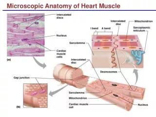
Cardiac Muscle • Striated, involuntary muscle • Found in walls of the heart • Consists of branching chains of stocky muscle cells. Uni- or binucleate. • Has sarcomeres & T-tubules • Cardiac muscle cells are joined by structures called intercalated discs – which consist of desmosomes and gap junctions. • Why do you suppose these are necessary? Notice the branching and the intercalated disc, indicated by the blue arrow.