Download

1 / 42
420 likes | 1.14k Views
Acute viral neurological diseases. Dr. Mohammed Arif Associate professor Consultant virologist Head of the virology unit College of medicine & KKUH. Acute viral neurological diseases. Aseptic meningitis Encephalitis Post-infectious encephalomyelitis Paralysis. 1-Aseptic meningitis.

E N D
Acute viral neurological diseases • Dr. Mohammed Arif • Associate professor • Consultant virologist • Head of the virology unit • College of medicine & KKUH
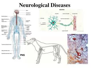
Acute viral neurological diseases • Aseptic meningitis • Encephalitis • Post-infectious encephalomyelitis • Paralysis
1-Aseptic meningitis • Case definition A syndrome characterized by acute onset of meningeal symptoms, fever and cerebrospinal fluid pleocytosis, with no laboratory evidence of bacterial or fungal meningitis.
Aseptic meningitis • Inflammation of the meninges that cover the brain and spinal cord. • Meninges: duramatter, arachnoid and piamatter. • It is a disease of children and young adults.
Viral etiology • Enteroviruses (85%). • Arboviruses. • Mumps virus. • Lymphocytic choriomeningitis. • Herpes viruses (HSV-1&2, CMV EBV.HHV-6,7,8)
Symptoms • Severe headache. • Fever. • Stiffness of neck. • Nausea. • Vomiting. • Sore throat. • Myalgia.
Symptoms ( CONT.) • Prognosis: recovery is usual in 5 – 14 days. • Complication : meningoencephalitis.
CSF-picture • Clear. • Colorless. • Pleocytosis (increased lymphocytes). • Increased protein. • Normal glucose. • Negative for bacterial culture.
2-Acute viral encephalitis • Inflammation of the brain. • Caused by direct viral invasion of the brain. • The virus replicate in the brain, causing destructive lesions in the grey matter.
Viral etiology • HSV-1&2. • Enteroviruses. • Arboviruses. • Rabies virus. • Mumps virus.
Clinical features • The incubation period 3-7 days. • Starts with the symptoms of aseptic meningitis followed by : • Drowsiness. • Mental confusion. • Alteration in the level of consciousness.
Clinical features (cont.) • Lack of coordination. • Seizures. • Coma.
Prognosis • The clinical outcome varies, some are mild with full recovery. • Others are severe with permanent neurological sequalae. • Permanent neurological impairments include memory, speech, vision, and muscle control.
Pathology • HSV-encephalitis is characterized by: • Edema, hemorrhage and necrosis confined primarily to the temporal lobes (bilateral or unilateral). • Vascular destruction with infiltrates of neutrophils and lymphocytes. • Glial proliferation. • Eosinophilic intranuclear inclusion bodies.
Pathology • Rabies encephalitis is characterized by: • Perivascular infiltrate of neutrophils and lymphocytes. • Glial proliferation. • Negri bodies, intracytoplasmic inclusion bodies, most prominent in the hippocampus.
Treatment • For enteroviruses and arboviruses, treatment is supportive. There is no anti-viral drug therapy. • For HSV, the recommended dose of acyclovir is 30 mg/ kg per day, in three divided doses for 14 - 21 days.
Methods of Diagnosis • Detection of the viral genome using PCR. • Isolation of the virus in tissue culture, followed by identification of the isolated virus. • MRI (magnetic resonance imaging) provides high quality picture of the brain, useful in the identification of edema, necrosis and hemorrhage.
Methods of Diagnosis (cont.) • CT-scan (computer assisted tomography), useful of identification of internal bleeding. Edema and necrosis. • EEG (electroencephalography) shows spikes and wave activity. Ewaves activity.
3-Post-infectious encephalomyelitis • It is uncommon complication of many viruses that do not usually affect the CNS including measles, mumps , rubella and varicella. • Encephalitis develops within 6 – 10 days after symptoms of viral illness appear.
Post- infectious encephalomyelitis • The disease is believed to be an auto-immune phenomenon, in which the immune system is triggered by exposure to foreign antigen which is closely related to host proteins normally expressed in brain tissue.
Pathology • Characterized by wide spread demyelinating lesions in the brain and spinal cord. • Lymphocytic infiltration and perivascular cuffing of adjacent blood vessels.
Clinical features • Similar to acute viral encephalitis. • Fever. • Headache. • Stiffness of neck. • Alteration in the level of consciousness. • Mental confusion. • Seizures and coma.
Prognosis • Not good. • Recovery is not complete. • Survivors are left with permanent neurological sequalae.
Treatment • Supportive therapy, there is no specific viral drug therapy.
Lab. diagnosis • By detection of the viral genome to measles, mumps and rubella. • By detection of IgM-Ab to these viruses.
Chronic viral neurological diseases • Subacute sclerosing pan encephalitis (SSPE). • Progressive multifocal leucoencephalopathy. • Tropical spastic paraparesis (TSP).
SSPE • Caused by a mutant measles virus. • Measles virus usually does not cause brain damage, but the mutant virus can invade the brain causing persistent infection and severe form of encephalitis and death. • It affects only unvaccinated children.
Clinical features • It is a rare, slowly progressive disease of the CNS ending in death. • SSPE develops 6 – 12 years after having a primary measles infection. • It is due to reactivation of persistent measles virus in the brain.
Clinical features • The disease starts with personality changes and memory defects followed by • Myoclonic. • Intellectual impairment. • Impairment of vision and speech. • Seizures. • Ataxia • Coma & death.
Laboratory Diagnosis • Presence of high Ab-titre to measles virus in CSF and serum. • MRI provides high quality picture of the brain. • Electro-encephalogram (ECG), is a test for the electrical activity of the brain, which shows a wave pattern whish is typical of SSPE.
Treatment • No cure for SSPE exist. • A combination of Isoprinosine and alpha interferon injected directly into the brain ventricles appears to be the most effective treatment.
Prevention • Immunization of children against measles virus is the only known prevention of SSPE.
Pathology • SSPE is characterized by atrophy of the cortex, loss of white matter and ventricular enlargement. • Perivascular lymphocytic infiltrate and intense gliosis. • Intranuclear inclusion bodies.
Progressive multifocal leukoencephalopathy • Caused by a polyoma virus known as JC virus. • The virus belongs to the family papovaviridae. • Genus: polyoma virus, • The virus is opportunistic, causing brain disorder only in the immunosuppressive patients. • Infection with JC virus is common, but asymptomatic. • 50 – 60% of adults have Ab to the virus.
Clinical features • It is a slowly progressive disease of the CNSthat is usually fatal. • It is due to reactivation of latent virus in the brain. • The disease is characterized by : Hemiparesis Dementia Impaired vision and speech Dysphagia Lack of coordination & seizures.
Prognosis • Death is very common 1 to 6 months when symptoms starts.
Laboratory diagnosis • Detection of the viral genome using PCR. • MRI.
Pathology • Brain autopsy shows multiple demyelination foci prominent in the hemispheres and cerebellum. • Destruction of the white matter. • Prominent gliosis. • Eosinophilic intranuclear inclusion bodies.