Download
1 / 35
370 likes | 729 Views
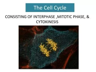
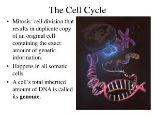
The Cell Cycle. Cell Division. Cell division – cell duplicates its DNA, separates the 2 copies and splits into 2 daughter cells Genome – entire collection of genetic information within the cell DNA is packaged into chromosomes. Human DNA Math for fun!.

E N D
Cell Division • Cell division – cell duplicates its DNA, separates the 2 copies and splits into 2 daughter cells • Genome – entire collection of genetic information within the cell • DNA is packaged into chromosomes
Human DNA Math for fun! • A single molecule of DNA has 1,000’s of genes! • A single strand of DNA ~5cm long • Each somatic cell has 46 molecules of DNA • 5cm x 46 = 230 cm of DNA per cell! • You have about 100 trillion cells • 230 cm x 100,000,000,000,000 = 23,000,000,000,000,000 cm of DNA in you body!!!!
How does it all fit? • 25mm capsule represents a prokaryotic cell (25,000x its actual size) • 17.5m of thread represents prokaryotic chromosome (25,000x its actual size)
How does it all fit? • 25mm capsule represents a prokaryotic cell (25,000x its actual size) • 17.5m of thread represents prokaryotic chromosome (25,000x its actual size) • SUPERCOILING!!
Chromosomes • Most prokaryotic cells have 1 long circular chromosome • Number of chromosomes in the nucleus of eukaryotic cells varies by species • Multicellular organisms have 2 types of cells: • Somatic cells – all body cells • 2 sets of chromosomes (1 from each parent) • Gametes – reproductive cells (sperm & egg) • 1 set of chromosomes
Eukaryotic Chromosomes • Gene: segment of DNA that codes for a protein or RNA molecule • DNA: stores information on which proteins to make, how, and when to make them • Chromatin: DNA and associated protein molecules called histones • Chromosome: condensed chromatin • Different chromosomes have varying amounts of genes
Gene Base Pair • Strand of DNA containing genes • DNA wraps itself around histones, making chromatin • Strand of chromatin with centromere • Chromatin duplicates itself during interphase (S), creating sister chromatids • Chromatids condense into chromosomes
Chromosome Duplication • Before cell division, each chromosome is duplicated • This results in 2 chromatids attached at the centromere
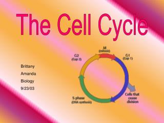
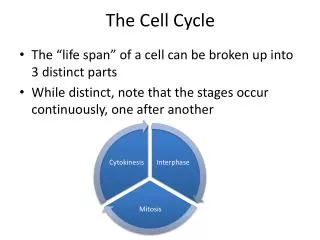
The Eukaryotic Cell Cycle • Cell Cycle: repeating sequence of cellular growth and division • Phases: • Interphase (90% of life): • G1 (1st growth phase) – cell grows and performs routine functions • S (Synthesis phase) – DNA is copied • G2 (2nd growth phase) – Microtubules are rearranged and cell prepares to divide • Mitosis– cell nucleus divides (each new nucleus has an identical copy of chromosomes) • Cytokinesis – cytoplasm divides • Results in 2 new identical cells
Cell Cycle… (IPMAT) • Interphase • G1 • S • G2 • Mitosis • Prophase • Metaphase • Anaphase • Telophase • Cytokinesis
Interphase • G1 • Growth and normal function • S • Chromatin duplicated (not yet visible) • G2 • 2 centrosomes form • Animal cells – each centrosome features 2 centrioles (microtubules arranged in a circle)
Prophase • Chromatin condenses into visible chromosomes • Nucleolus disappears • Centrosomes move to opposite poles of cell • Nuclear envelope fragments
Metaphase • Centrosomes are at opposite ends of the cell • Chromosomes line up at midline of cell • Each kinetochore has a microtubule that extends to the centrosome
Anaphase • 2 sister chromatids separate (becoming a chromosome) • Chromatids move to opposite poles as microtubules shorten • Cell elongates • By the end, the two ends of the cell each have a identical set of chromosomes
Telophase • Nuclear envelopes reform • Chromosomes become less condensed
Cytokinesis - Animal Cells • Cleavage furrow forms and pinches the cell into 2
Cytokinesis - Plant Cells • Vesicles fuse at midline to form cell plate; cell wall forms from cell plate separating the 2 new plant cells
Challenge Question • Explain the role of microtubules in animal cell division • Hint: MTOC Mitosis Cleavage Furrow • Pg 246-247
Prokaryotic Cell Division • Binary Fission: divides into 2 new daughter cells
Do Now: • What would be most useful to know about cancer? • What are you most curious about? • Why is it important to become educated about health issues and prevention?
Cell Cycle Control System • The timing and rate of cell division in various parts of an organism are crucial to normal growth, development, and maintenance • Molecules within the cell trigger and coordinate key events of the cell cycle • Both internal and external factors can affect cell cycling
Checkpoints • Checkpoint – critical control point where “stop” and “go” signals regulate the cycle (inspection station) • 3 major checkpoints: • G1 (Most important in animal cells) • Is this a dividing cell? • Is this cell healthy? • Is this cell large enough? • **If cell does not receive the “go” signal, it will switch into G0 non-dividing phase • G2 • Is DNA copied properly? • M • Restarts cell cycle to G1 phase
Cancer • At the checkpoints • Oncogenes – tell cell cycle to “go” • Tumor Suppressor Genes – tell cell cycle to “stop” • If either of the genes that control the cell cycle become mutated • Mutagens – cause mutations • Carcinogens – mutagens cause cancer • The cell cycle goes too fast and divides out of control This is called cancer. • Cells pile up in one area and form a tumor • Immune system does not recognize as foreign • Benign tumor – does not invade adjacent tissue • Malignant Tumor – cancer spreads and new tumors form (metastasize)
Pathology of Cancer Tumor Grade Cancer Stage 0. abnormal cells (pre-cancerous) 2cm or less 2-5 cm 5cm Metastatic – spread throughout body • Similar to healthy cells • Different from healthy cells • Very different from healthy cells • Vastly different from healthy cells