Download
1 / 19
200 likes | 401 Views
Unusual Optic Nerve Sheath Meningioma (Kennerdell Case 12). John S. Kennerdell, MD Chair, Department of Ophthalmology Allegheny General Hospital Professor, Ophthalmology Drexel University College of Medicine. Case Presentation.

E N D
Unusual Optic Nerve Sheath Meningioma (Kennerdell Case 12) John S. Kennerdell, MD Chair, Department of Ophthalmology Allegheny General Hospital Professor, Ophthalmology Drexel University College of Medicine
Case Presentation • 39-year-old female c/o 6 year history of blurred vision OS, Darkening of vision in eccentric gaze OS, dark lines in vision OS • Vision: 20/20 OD 20/25 OS 9/1996 • Ishihara: 12/12 OD 10/12 OS • Pupils: 3/3 RRL 2+ RAPD OS • Motility: 2+ abduction deficit OS, 8∆ esotropia, 20∆ esotropia L gaze Unusual Optic Nerve Sheath MeningiomaSlide 1/18
Case Presentation • Exophthalmometry: No proptosis • SLE: Normal • DFE: OD: Disc Normal OS: Moderate chronic disc edema • Macula/Retina: OD: Normal OS: Choroidal folds Unusual Optic Nerve Sheath MeningiomaSlide 2/18
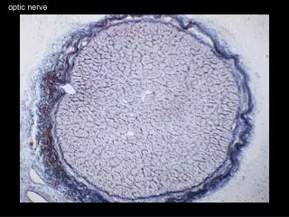
An enlarged blind spot caused by the meningioma choking the left optic nerve. Unusual Optic Nerve Sheath MeningiomaSlide 3/18
The left orbital meningioma non-contrast scan (right on this slide). Unusual Optic Nerve Sheath MeningiomaSlide 4/18
The coronal view of the enhancing meningioma (right on this slide). Unusual Optic Nerve Sheath MeningiomaSlide 5/18
Treatment • 10/7/96 to 11/13/96 Stereotactic XRT 28 fractions 5040 cGy Intraorbital and intracranial ON 4MV Ph Unusual Optic Nerve Sheath MeningiomaSlide 6/18
Clinical Followup • 8/1997: Followup Exam: 20/20 OU “Disc pale”, choroidal folds • 5/1998: C/O enlarging scotoma, dim vision 20/20 OD 20/200 OS 0/12 plates Persistent RAPD OS Severe disc edema with macular and peripapillary exudates Unusual Optic Nerve Sheath MeningiomaSlide 7/18
Clinical Followup • 9/1998: Persistent poor vision 20/20 OD, 20/200 OS Florid disc edema with macular exudates OS, choroidal folds • MRI repeated, no radiographic progression no new enhancement Unusual Optic Nerve Sheath MeningiomaSlide 8/18
The severe disc edema that recurred. Unusual Optic Nerve Sheath MeningiomaSlide 9/18
The severe central visual field defect of the left eye. Unusual Optic Nerve Sheath MeningiomaSlide 10/18
The enhancing left optic nerve meningioma lesion with apparent fluid distal to the lesion abutting the globe (right side of slide). Unusual Optic Nerve Sheath MeningiomaSlide 11/18
Surgical Therapy • 9/17/98 Left lateral optic nerve sheath biopsy and fenestration • 6 hours post op: 20/20 OD, NLP OS Pupil: dilated, nonreactive • Hospitalized for observation solumedrol 250mg IV q6, 80mg pred taper • POD #1: 20/20 OD, 10/400 OS Unusual Optic Nerve Sheath MeningiomaSlide 12/18
The lateral approach to the lesion without removing the left orbital rim. Unusual Optic Nerve Sheath MeningiomaSlide 13/18
Post Operative Followup • POW #5: 20/20 OD, 20/25 OS Persistent florid edema, macular exudates OS Unusual Optic Nerve Sheath MeningiomaSlide 14/18
The post operative appearance of the lesion which is much smaller now. Note the slight adhesion of the optic nerve lesion to the lateral orbitotomy site (right on this slide). Unusual Optic Nerve Sheath MeningiomaSlide 15/18
The post operative appearance of the patient. Unusual Optic Nerve Sheath MeningiomaSlide 16/18
The marked improvement of the visual field with the vision returning again to 20/20. Unusual Optic Nerve Sheath MeningiomaSlide 17/18
The marked reduction in temporal edema following the left optic nerve sheath decompression anterior to the lesion. Unusual Optic Nerve Sheath MeningiomaSlide 18/18