Download
1 / 91
910 likes | 921 Views
Welcome to Anatomy II! This lecture covers the changes in the course compared to last year's Anatomy I, including attendance taken in lecture and lab, implementation of "mastering anatomy" homework, and study tips. The lecture then provides an overview of blood components, including red blood cells and hemoglobin, regulation of blood components, blood typing, and hemostasis. It also introduces the cardiovascular system and its functions.

E N D
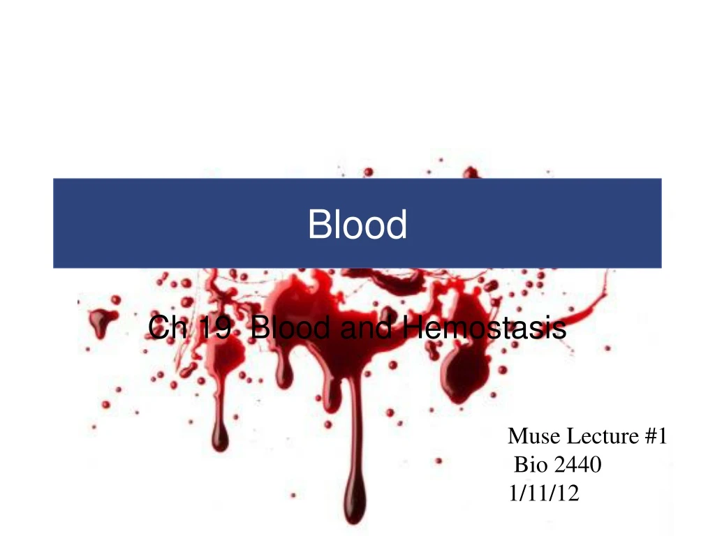
Blood Ch 19 Blood and Hemostasis Muse Lecture #1 Bio 2440 1/11/12
Welcome to Anatomy II Changes in course compared to last year’s Anatomy I - attendance taken in lecture and lab- make sure you sign the attendance sheet. - implementation of “mastering anatomy” homework (20%) Study Tips- bookmark website - outline as you read - attend SI - assess before the test - evaluate study groups - use all of your lab time - get questions answered
Ch 19 Overview Blood components Red Blood cells and hemoglobin Regulation of Blood components Blood typing Hemostasis
Introduction to the Cardiovascular System • A circulating transport system • A pump (the heart) • A conducting system (blood vessels) • A fluid medium (blood) • Is specialized fluid of connective tissue • Contains cells suspended in a fluid matrix
Introduction to the Cardiovascular System • To transport materials to and from cells • Oxygen and carbon dioxide • Nutrients • Hormones • Immune system components • Waste products
Functions of Blood • Transport of dissolved substances • Regulation of pH and ions • Restriction of fluid losses at injury sites • Defense against toxins and pathogens • Stabilization of body temperature
Physical Characteristics of Blood • Whole Blood • Plasma • Fluid consisting of: • water • dissolved plasma proteins (albumins and globulins) • other solutes (salt, dissolved gasses) • Formed elements • All cells and solids
Physical Characteristics of Blood Figure 19–1 The Composition of Whole Blood
Physical Characteristics of Blood Figure 19–1b The Composition of a Typical Sample of Plasma
Physical Characteristics of Blood Figure 19–1c The Composition of Formed Elements of Blood
Physical Characteristics of Blood • Three Types of Formed Elements • Red blood cells(RBCs) or erythrocytes • Transport oxygen - red because of hemoglobin • White blood cells(WBCs) or leukocytes • Part of the immune system • Platelets • Cell fragments involved in clotting
Physical Characteristics of Blood • Hemopoiesis • Process of producing formed elements • By myeloid and lymphoid stem cells • Fractionation • Process of separating whole blood for clinical analysis • Into plasma and formed elements centrifugation or filtering
Physical Characteristics of Blood • Three General Characteristics of Blood • 38°C (100.4°F) is normal temperature • High viscosity • Slightly alkaline pH (7.35–7.45)
Physical Characteristics of Blood • Blood volume (liters) = 7% of body weight (kilograms) • Adult male: 5 to 6 liters • Adult female: 4 to 5 liters
Plasma • Makes up 50–60% of blood volume • More than 90% of plasma is water • Extracellular fluids • Interstitial fluid (IF) and plasma • Materials plasma and IF exchange across capillary walls • Water • Ions • Small solutes
Plasma • Differences between Plasma and IF • Levels of O2 and CO2 • Concentrations and types of dissolved proteins • Plasma proteins do not pass through capillary walls
Plasma • Plasma Proteins • Albumins (60%) • Transport substances such as fatty acids, thyroid hormones, and steroid hormones. HSA- Human Serum Albumin is also a redox buffer to protect proteins from oxidation. • Globulins (35%) • Antibodies, also called immunoglobulins • Transport globulins (small molecules): hormone-binding proteins, metalloproteins, apolipoproteins (lipoproteins), and steroid-binding proteins • Fibrinogen (4%) • Molecules that form clots and produce long, insoluble strands of fibrin
Plasma • Serum • Liquid part of a blood sample • In which dissolved fibrinogen has converted to solid fibrin • Other Plasma Proteins • 1% of plasma • Changing quantities of specialized plasma proteins • Enzymes, hormones, and prohormones
Plasma • Origins of Plasma Proteins • 90% + made in liver • Antibodies made by plasma cells (WBCs(B-cells) • Peptide hormones made by endocrine organs
Red Blood Cells • Red blood cells (RBCs) make up 99.9% of blood’s formed elements • Hemoglobin • The red pigment that gives whole blood its color • Binds and transports both oxygen and carbon dioxide
Red Blood Cells • Abundance of RBCs • Red blood cell count: the number of RBCs in 1 microliter of whole blood • Male: 4.5–6.3 million • Female: 4.2–5.5 million • Hematocrit (packed cell volume, PCV): percentage of RBCs in centrifuged whole blood • Male: 40–54 • Female: 37–47
Red Blood Cells • Structure of RBCs - anucleate in mammals • Small and highly specialized discs • Thin in middle and thicker at edge • Importance of RBC Shape and Size • High surface-to-volume ratio • Quickly absorbs and releases oxygen • Discs form stacks called rouleaux • Smooth the flow through narrow blood vessels • Discs bend and flex entering small capillaries: • 7.8 µm RBC passes through 4 µm capillary Figure 19–2d
Red Blood Cells Figure 19–2a–c The Anatomy of Red Blood Cells
Red Blood Cells Figure 19–2d The Anatomy of Red Blood Cells
Red Blood Cells • Lifespan of RBCs • Lack nuclei, mitochondria, and ribosomes • Means no repair and anaerobic metabolism • Live about 120 days
Red Blood Cells • Hemoglobin (Hb) • Protein molecule, that transports respiratory gases • Normal hemoglobin (adult male) • 14–18 g/dL whole blood • Normal hemoglobin (adult female) • 12–16 g/dL, whole blood
Red Blood Cells • Hemoglobin Structure • Complex quaternary structure • Four globular protein subunits: • Each with one molecule of heme • Each heme contains one iron ion • Iron ions • Associate easily with oxygen (oxyhemoglobin) • OR • Dissociate easily from oxygen (deoxyhemoglobin) Figure 19–3
Red Blood Cells Figure 19–3 The Structure of Hemoglobin
bGlobin chains Heme group a Globin chains (a) Hemoglobin consists of globin (two alpha and two beta polypeptide chains) and four heme groups. (b) Iron-containing heme pigment. Figure 17.4
Red Blood Cells • Fetal Hemoglobin • Strong form of hemoglobin found in embryos • Takes oxygen from mother’s hemoglobin
Red Blood Cells • Hemoglobin Function • Carries oxygen • With low oxygen (peripheral capillaries) • Hemoglobin releases oxygen • Binds carbon dioxide and carries it to lungs • Forms carbaminohemoglobin
Red Blood Cells Figure 19–4 ”Sickling” in Red Blood Cells B-globin D6V
Red Blood Cells • RBC Formation and Turnover • 1% of circulating RBCs wear out per day • About 3 million RBCs per second • Macrophages of liver, spleen, and bone marrow • Monitor RBCs • Engulf RBCs before membranes rupture (hemolyze)
Red Blood Cells • Hemoglobin Conversion and Recycling • Phagocytes break hemoglobin into components • Globular proteins to amino acids • Heme to biliverdin • Iron • Hemoglobinuria • Hemoglobin breakdown products in urine due to excess hemolysis in bloodstream • Hematuria • Whole red blood cells in urine due to kidney or tissue damage
Red Blood Cells • Iron Recycling • Iron removed from heme leaving biliverdin • To transport proteins (transferrin) • To storage proteins (ferritin and hemosiderin)
Red Blood Cells • Breakdown of Biliverdin • Biliverdin (green) is converted to bilirubin (yellow) • Bilirubin is: • excreted by liver (bile) • jaundice is caused by bilirubin buildup • converted by intestinal bacteria to urobilins and stercobilins
Red Blood Cells Figure 19–5 Recycling of Red Blood Cell Components
Red Blood Cells • RBC Production • Erythropoiesis • Occurs only in myeloid tissue (red bone marrow) in adults • Stem cells mature to become RBCs • Hemocytoblasts • Stem cells in myeloid tissue divide to produce • Myeloid stem cells: become RBCs, some WBCs • Lymphoid stem cells: become lymphocytes
Red Blood Cells • Stages of RBC Maturation • Myeloid stem cell • Proerythroblast • Erythroblasts • Reticulocyte • Mature RBC
Red Blood Cells Figure 19–6 Stages of RBC Maturation
Enters blood stream Stem cell Committed cell Developmental pathway Phase 1 Ribosome synthesis Phase 2 Hemoglobin accumulation Phase 3 Ejection of nucleus Reticulo- cyte Erythro- cyte Proerythro- blast Early erythroblast Late erythroblast Normoblast Hemocytoblast Figure 17.5
Red Blood Cells • Regulation of Erythropoiesis • Building red blood cells requires • Amino acids • Iron • Vitamins B12, B6, and folic acid: • pernicious anemia • low RBC production • due to unavailability of vitamin B12
This is why athletes train in low altitude IMBALANCE Homeostasis: Normal blood oxygen levels 1 Stimulus: Hypoxia (low blood O2- carrying ability) due to • DecreasedRBC count • Decreased amountof hemoglobin • Decreasedavailability of O2 5 O2- carryingability of bloodincreases. IMBALANCE 4 Enhancederythropoiesisincreases RBCcount. 2 Kidney (and liver toa smaller extent)releaseserythropoietin. 3 Erythropoietinstimulates redbone marrow. Figure 17.6
Red Blood Cells • Stimulating Hormones • Erythropoietin (EPO) • Also called erythropoiesis-stimulating hormone • Secreted when oxygen in peripheral tissues is low (hypoxia) • Due to disease or high altitude
Blood Typing • Are cell surface proteins that identify cells to immune system • Normal cells are ignored and foreign cells attacked • Blood types • Are genetically determined • By presence or absence of RBC surface antigens A, B, Rh (or D)
Blood Typing Landsteiner • Four Basic Blood Types • A (surface antigen A) • B (surface antigen B) • AB (antigens A and B) • O (neither A nor B)
Blood Typing Figure 19–7a Blood Types and Cross-Reactions
Blood Typing • Agglutinogens • Antigens on surface of RBCs • Screened by immune system • Plasma antibodies attack and agglutinate (clump) foreign antigens
Blood Typing • Blood Plasma Antibodies • Type A person • Type B antibodies in sera • Type B • Type A antibodies in sera • Type O • Both A and B antibodies in sera • Type AB • Neither A nor B antibodies in sera