Download
1 / 21
240 likes | 272 Views
CHAPTER 27 Reproduction and Embryonic Development. Modules 27.9 – 27.15. PRINCIPLES OF EMBRYONIC DEVELOPMENT. 27.9 Fertilization results in a zygote and triggers embryonic development. The shape of a human sperm cell is adapted to its function. Middle piece. Neck. Head. Plasma membrane.

E N D
CHAPTER 27Reproduction and Embryonic Development Modules 27.9 – 27.15
PRINCIPLES OF EMBRYONIC DEVELOPMENT 27.9 Fertilization results in a zygote and triggers embryonic development • The shape of a human sperm cell is adapted to its function Middlepiece Neck Head Plasma membrane Tail Mitochondrion(spiral shape) Nucleus Acrosome Figure 27.9B
Only one of these sperm will penetrate this human egg cell to initiate fertilization • Fertilization is the union of a sperm and an egg to form a diploid zygote Figure 27.9A
1 The spermapproachesthe egg 2 The sperm’sacrosomal enzymesdigest the egg’s jelly coat 3 Proteins on thesperm head bindto egg receptors SPERM 4 The plasma membranesof sperm and egg fuse • Process of fertilization Spermhead The spermnucleus enters the egg cytoplasm 5 Nucleus Acrosome Acrosomalenzymes Plasmamembrane Afertilizationenvelopeforms 6 Receptor protein molecules Plasmamembrane Spermnucleus Vitellinelayer Cytoplasm Eggnucleus Jellycoat The nucleiof spermand egg fuse 7 EGG CELL Zygotenucleus Figure 27.9C
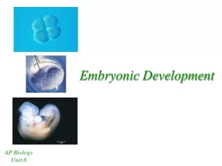
27.10 Cleavage produces a ball of cells from the zygote • Cleavage is the first major phase of embryonic development • It is the rapid succession of cell divisions • It creates a multicellular embryo from the zygote • It partitions the multicellular embryo into developmental regions
ZYGOTE 2 cells • Cleavage in a sea urchin 4 cells 8 cells Blastocoel Many cells(solid ball) BLASTULA(hollow ball) Cross sectionof blastula Figure 27.10
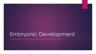
27.11 Gastrulation produces a three-layered embryo • Gastrulation is the second major phase of embryonic development • It adds more cells to the embryo • It sorts all cells into three distinct cell layers • The embryo is transformed from the blastula into the gastrula
Ectoderm, the outer layer • Endoderm, an embryonic digestive tract • Mesoderm, which partly fills the space between the ectoderm and endoderm • The three layers produced in gastrulation
Animal pole Blastocoel 1 Vegetal pole BLASTULA • Development of frog gastrula GASTRULATION 2 Blastoporeforming Blastoporeforming Blastocoelshrinking Archenteron 3 Archenteron Ectoderm Mesoderm 4 Endoderm Yolk plug Yolk plug GASTRULA Figure 27.11C
27.12 Organs start to form after gastrulation • Embryonic tissue layers begin to differentiate into specific tissues and organ systems • In chordates • the notochord develops from the mesoderm • the neural tube develops from the ectoderm • The neural tube becomes the brain and spinal cord
Neuralplate Neuralfold Neuralfold Neuralplate Notochord Ectoderm Mesoderm Endoderm Archenteron Neural folds Outer layerof ectoderm Neural tube Figure 27.12A, B
Neural tube Notochord Somite Coelom • Somites are blocks of mesoderm that will give rise to segmental structures Archenteron(digestive cavity) Somites Tail bud • The body cavity, or coelom, also develops from the mesoderm Eye Figure 27.12C
The tissues and organs of a tadpole emerge from cells of the ectoderm, mesoderm, and endoderm Figure 27.12D
27.13 Changes in cell shape, cell migration, and programmed cell death give form to the developing animal Ectoderm • Tissues and organs take shape in a developing embryo as a result of • cell shape changes • cell migration Figure 27.13A
Cellsuicide Dead cellengulfed anddigested byadjacentcell • programmed cell death (apoptosis) Figure 27.13B
27.14 Embryonic induction initiates organ formation • Induction is the mechanism by which one group of cells influences the development of tissues and organs from ectoderm, endoderm, and mesoderm • Adjacent cells and cell layers use chemical signals to influence differentiation • Chemical signals turn on a set of genes whose expression makes the receiving cells differentiate into a specific tissue
Induction during egg development Optic cup Lens ectoderm Cornea Futurebrain Lens Opticvesicle Futureretina Opticstalk 1 2 3 4 Figure 27.14
27.15 Pattern formation organizes the animal body • Pattern formation is the emergence of a body form with structures in their correct relative positions • It involves the response of genes to spatial variations of chemicals in the embryo
Wing development ANTERIOR Birdembryo VENTRAL Normal wing Limb bud DISTAL Limb bud develops DORSAL PROXIMAL POSTERIOR Figure 27.15A
Pattern-forming zone Wing withduplication Graft of cellsfrom pattern-formingzone Graft Donorlimbbud Hostlimbbud Host limb bud develops Host pattern-forming zone Donor cells Figure 27.15B