Download
1 / 20
210 likes | 220 Views
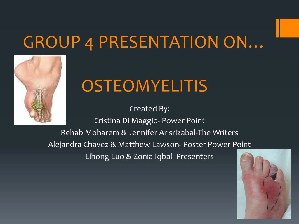
GROUP 4 PRESENTATION ON… OSTEOMYELITIS. Created By: Cristina Di Maggio- Power Point Rehab Moharem & Jennifer Arisrizabal-The Writers Alejandra Chavez & Matthew Lawson- Poster Power Point Lihong Luo & Zonia Iqbal- Presenters. BRIEF PATIENT HISTORY.

E N D
GROUP 4 PRESENTATION ON…OSTEOMYELITIS Created By: Cristina Di Maggio- Power Point Rehab Moharem & Jennifer Arisrizabal-The Writers Alejandra Chavez & Matthew Lawson- Poster Power Point Lihong Luo & Zonia Iqbal- Presenters
BRIEF PATIENT HISTORY A 49 year old male patient was transported to the Emergency Room at Lutheran Medical Center. The patient was an in patient that was brought in on a stretcher, therefore he was non-ambulatory. The patient was previously diagnosed with Osteomyelitis which led to an amputation of the 5th distal phalanx, middle phalanx, and metatarsal of the patient’s right foot. A right foot order was placed to rule out the spread of Osteomyelitis to any other bones of the patient’s right foot.
VIEWS ORDERED BY PHYSICIAN The projections ordered by the patient’s doctor was an AP view, an Oblique view, and a Lateral view of the patient’s right foot; and due to the condition of the patient’s foot the radiographs were taken directly from the stretcher. For the AP view the patient’s right foot was positioned flat and parallel to the IR and so that the central ray entered 10-15 degrees cephalic to the base of the 3rd metatarsal; this view was taken at 62kV with 1.6mAS. For the Oblique view the patient’s right root was internally rotated 30 degrees and positioned so that the central ray entered perpendicular to the base of the 3rd metatarsal; this view was taken at 62kV with 2mAS. For the Lateral view the patient’s right foot was turned to rest on the lateral aspect and positioned so the central ray entered perpendicular the base of the 3rd metatarsal; this view was taken at 64kV with 2.4mAS.
NO contrast media was administered to the patient for this exam.
ANATOMY & PHYSIOLOGY • The foot is made up of 26 bones as well as joints, muscles, and soft tissue. • The foot is also divided into 3 categories…the forefoot, the midfoot, and the hindfoot • The forefoot consists of the phalanges & metatarsals… the midfoot contains the tarsals….and the hindfoot contains the talus and calcaneus.
WHAT IS OSTEOMYELITIS & HOW IT CAN OCCUR Osteomyelitis is an infection and inflammation within a bone. The infection can occur in a few different ways: 1 - it can either occur internally via the blood stream; for example if somebody has an pneumonia, that can spread to the bone through our bloodstream causing that bone to become infected. 2- it can also occur externally; for example if a person has an open wound or a tissue infection, that can spread directly to the bone causing the bone to become infected.
Example of Osteomyelitis occuring via open wound
Example of Osteomyelitis occurring via bloodstream
STAGES OF OSTEOMYELITITS Stage 1 is known as Acute Osteomyelitis; Acute Osteomyelitis is when the infection to the bone develops within the first 2 weeks of an injury or the start of an ongoing disease. Stage 2 is known as Subacute Osteomyelitis; Subacute Osteomyelitits is when infection to the bone develops within 1 or 2 months of an injury or the start of an ongoing disease. Stage 3 is known as Chronic Osteomyelitis; Chronic Osteomyelitis is when infection to the bone develops at least 2 months after an injury or the start of an ongoing disease. Osteomyelitis is categorized into 3 stages
HOW OSTEOMYELITIS IS DETECTED There are 3 tests that can be done to diagnose Osteomyelitis. The 3 tests are: a Blood test, a bone biopsy, and an imaging test. Imaging tests that can be done are either conventional x-ray, CT, or MRI…in our patients’ case conventional x-ray was chosen. It is common for a doctor to order these certain tests and run procedures to diagnose Osteomyelitis. These tests will aid in helping to determine which germ is causing the infection.
TREATMENTS FOR OSTEOMYELITIS If the infection to the bone is at a mild state, antibiotics can be prescribed to destroy the bacteria before the infection gets worse. When the infection to the bone is at a more severe state…surgery may be needed to remove dead bone tissue therefore resulting in an amputation To the right is the AP view of our patients’ right foot…as you can see the distal phalanx, middle phalanx and metatarsal of the patient’s 5th digit was amputated due to the infection; therefore his infection was at a more severe state.
The amputation of the patient’s 5th digit can also be seen in the Oblique projection of the patient’s right foot
BEFORE AND AFTER AMPUTATION This is an Oblique view of another patient’s foot. This patient also suffered from Osteomyelitis, but of the 4th metatarsal…this image was taken prior to the amputation. This is the AP view of our patient’s foot with Osteomyelitis of the 5th metatarsal…and this image was taken after the amputation of the patient’s distal phalanx, middle phalanx, and metatarsal.
WHO IS AT RISK? Osteomyelitis can occur in people who experience the following: 1- weakened immune systems 2- circulation problems 3- recent trauma 4-diabetes 5- patients on dialysis 6- sometimes even patients with urinary catheters Osteomyelitis can affect anyone from infants and children to adults and the elderly.
Patients with weakened immune Systems are more susceptible to the following… therefore are at a greater risk of acquiring Osteomyelitis. Patients experiencing poor circulation are more susceptible to acquiring Osteomyelitis because if not enough blood flows through the body, that can cause tissues to die as well as bones to become infected.
HOW TO PREVENT OSTEOMYELITIS There are a few ways Osteomyelitis can be prevented… 1- Taking certain measures to avoid cuts, wounds, or scrapes. By doing so, this will prevent germs from entering the body and potentially causing an infection. 2- If it does occur that you get a cut, wound, or scrape make sure to properly disinfect the area and apply a clean bandage. 3- DON’T FORGET to frequently check the wound for signs of infection
IN CONCLUSION… Today, not only did we learn what Osteomyelitis is…we also learned a few things that come along with it. For example we learned how Osteomyelitis can occur, the different stages it has, how Osteomyelitis can be detected, how it can be treated, who is at risk, and also how it can be prevented…So next time you get a “little scrape” or just “a cold” make sure you properly treat it…or you can end up like this
REFERENCES • Eugene D. Frank, Bruce W. Long, Barbara J. Smith. Merrill’s Atlas of Radiographic Positioning and Procedures: Volume 1, 12e. March 2, 2011. Print. • Davis BL. Kuznicki J. Praveen SS. Sferra JJ. Lower-extremity amputations in patients with diabetes: pre-and post- surgical decisions related to successful rehabilitation. Diabetes Metab Res Rev 2004: Web 2 October 20013. • Kofler, Johann. Feist Melanie. Starke, Alexander. Nuss,Karl. Berliner Munchener Tieraztliche Wochenschift Volume:120. P56-164.published:Mar-Apr 2007. Web. 2 October 2013. • Malizos, Konstantions N.; Gougoulias, Nikolaos E.; DAILIANA, Zoe H.; Varitimidis, Sokrati; Bargiotas, Konstantinos A.; Paridis, Dionysios. Injury. Mar 2010, Vol. 41 Issue 3, p285-293. Web. 2 October 2013.