Download
1 / 26
260 likes | 275 Views
Explore detailed information on the axial skeleton, skull structure, and vertebral anatomy including bones, sutures, foramina, and processes. Understand the intricate details of the human skull and spinal column.

E N D
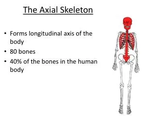
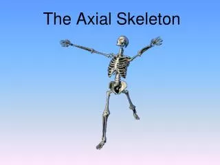
The Axial Skeleton Study Guide
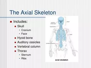
right side of the skull Frontal bone Coronal suture Sphenoid bone Parietal bone Squamous suture Ethmoid bone Lacrimal bone Lambdoid suture Lacrimal fossa Occipital bone Nasal bone Temporal bone Zygomatic process Zygomatic bone Maxilla External acoustic meatus Mastoid process Styloid process Mandible Condylar process Mental foramen Mandibular notch Coronoid process Mandibular ramus Mandibularangle
Frontal bone Parietal bone Supraorbital foramen Nasal bone Sphenoid bone Superior orbital fissure Temporal bone Optic canal Ethmoid bone Inferior orbital fissure Lacrimal bone Zygomatic bone Infraorbital foramen Maxilla Vomer Mandible Mentalforamen
Parietal bone Sagittal suture Lambdoidsuture Occipital bone Superior nuchal line External occipitalprotuberance Inferior nuchal line Occipital condyle
Maxilla Hardpalate Palatine bone Infraorbital foramen Maxilla Sphenoid bone Zygomatic bone Vomer zygomatic process External acoustic meatus Styloid process Mastoid process Jugular foramen Temporal bone Occipital condyle Occipital bone External occipitalprotuberance Foramen magnum Inferior view of the skull (mandible removed)
The Holes in the Head Optic foramen Optic fissure Foramen rotundum Foramen ovale Foramen spinosum Foramen lacerum Internal acoustic meatus Jugular foramen Hypoglossal canal Foramen magnum ▲
Crista galli Ethmoidbone View Cribriform plate Frontal bone Optic canal Lesser wing Sphenoid Greater wing Foramen rotundum Foramen ovale sella turcica Foramen spinosum Foramen lacerum Internal acousticmeatus Temporal bone Jugular foramen Hypoglossal canal Parietal bone Occipital bone Foramen magnum Superior view of the skull, calvaria removed
View Superior view of the skull, calvaria removed
Parietal bone Coronal suture Frontal bone Sphenoid bone Squamoussuture Temporal bone Crista galli Lambdoid suture Nasal bone Occipital bone Ethmoid bone Vomer External occipitalprotuberance Maxilla Internal acousticmeatus Mandible Mandibularforamen Palatine bone Midsagittal section showing the internal anatomy of the left half of skull
Midsagittal section showing the internal anatomy of the left half of skull
Mandible Temporomandibularjoint Coronoidprocess Mandibular notch Condylarprocess Mandibular foramen Ramusofmandible Mental foramen Mandibularangle Body of mandible Mandible, right lateral view
Mandible Mandible, right lateral view
IX. Structure of a Vertebra Posterior Lamina Spinousprocess Transverseprocess Superiorarticularprocessandfacet Vertebralforamen Pedicle Body Anterior
Posterior Anterior
Posterior view Anterior view
Sacral promontory Body Facet of superiorarticular process Sacralcanal Ala Body offirstsacralvertebra Auricularsurface Mediansacralcrest Lateralsacral crest Transverse ridges(sites of vertebralfusion) Posteriorsacralforamina Anteriorsacralforamina Apex Sacralhiatus Coccyx Coccyx Posterior view Anterior view
XIII. Thoracic Cage Jugular notch Clavicular notch Manubrium Sternal angle Body Sternum Trueribs(17) Xiphoidprocess Falseribs(812) Costalcartilage L1Vertebra Floatingribs (11, 12)
XIII. Thoracic Cage L1Vertebra
Superior costal facet(for head of rib) Angleof rib Body of vertebra Head of rib Intervertebral disc Neck of rib Tubercle of rib Shaft Sternum Cross-sectionof rib Costal cartilage Vertebral and sternal articulations of a typical true rib
Sternum Cross-sectionof rib Vertebral and sternal articulations of a typical true rib
Figure 7.25c Ribs. Articular faceton tubercle of rib Spinous process Shaft Transversecostal facet(for tubercleof rib) Ligaments Neck of rib Body ofthoracicvertebra Head of rib Superior view of the articulation between a rib and a thoracic vertebra
Figure 7.25c Ribs. Superior view of the articulation between a rib and a thoracic vertebra