Download
1 / 76
770 likes | 1.17k Views
Kontrol Terhadap Fungsi Pencernaan. dr. Nuraiza Meutia,M.Biomed Departemen Fisiologi FK USU. Aktivitas saluran cerna untuk menjalankan proses percernaan diatur oleh sistem saraf dan endokrin. Saluran cerna memiliki kemandirian untuk kedua sistem tersebut.

E N D
Kontrol Terhadap Fungsi Pencernaan dr. Nuraiza Meutia,M.Biomed Departemen Fisiologi FK USU
Aktivitas saluran cerna untuk menjalankan proses percernaan diatur oleh sistem saraf dan endokrin. • Saluran cerna memiliki kemandirian untuk kedua sistem tersebut. • Terjadi aktivitas terintegrasi antara kedua sistem mengatur aktivitas motorik dan sekretorik
Pada akhir perkuliahan, anda harus dapat : • Menjelaskan anatomi fungsional dinding GIT • Menjelaskan aktivitas utama GIT • Menjelaskan persarafan otonom yang mengatur GIT • Menjelaskan letak dan fungsi pleksus saraf di GIT • Menyebutkan nama dan efek neurotransmitter yang bekerja di ENS • Menjelaskan peran hormon dalam kontrol GIT (nama hormon, sumber, target dan efek) • Menjelaskan proses integrasi sistem saraf dan endokrin di GIT • Menjelaskan perbedaan refleks lokal dan refleks sentral dalam regulasi kerja GIT • Menjelaskan aktivitas listrik pada otot polos GIT • Menjelaskan mekanisme kontraksi dan jenis motilitas GIT • Menjelaskan contoh-contoh refleks GI • Menjelaskan regulasi aktivitas lambung, usus halus, usus besar, pankreas dan kandung empedu.
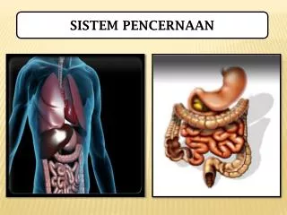
Digestive Process • Aktivitas GIT : • Motilitas • Sekresi • Digesti • Absorbsi
Fungsi sistem regulasi di GIT : • Mengatur aktivitas motilitas dan sekresi • Mengatur aliran darah ke GIT • Menerima dan menyampaikan informasi melalui neuron sensori (aferen) , dari reseptor-reseptor yang menerima stimulus mekanikal, thermal, osmotik dan kimiawi.
Aktivitas GIT diatur oleh sistem Saraf Otonom Terdiri dari : - Divisi parasimpatetik - Divisi simpatetik - Enteric Nervous System (ENS)
Parasimpatetik N. Vagus & N.pelvik Neuron preganglionik panjang; postganglionik pendek, bersinaps dengan neuron ENS Stimulasi eksitasi aktivitas ENS Mengandung serat sensori aferen (80 %) N.Vagusbersinaps ke neuron ENS di esophagus, lambung,usus halus, sebagian kolon, kandung empedu, & pankreas N.Pelvik bersinaps dengan ENS di usus besar Neurotransmitter : Ach
Simpatetik Serat simpatetik ke GIT berasal dari medula spinalis segmen T-5 sampai L-2. Neurotransmitter : norepinefrin Aktivitas simpatetik inhibisi motilitas dan sekresi GIT, konstriksi sfinkter dan pembuluh darah.
Enteric Nervous System Enteric Nervous System (ENS) terdapat di seluruh dinding GIT, mulai esophagus sampai anus. Terbentuk dari 100 juta neuron (mengimbangi spinal cord). Memiliki 3 jenis neuron : sensori, motorik, & interneuron ENStersusun atas 2 pleksus utama : (1) MyentericplexusatauAuerbach'sPlexus: berada di antara lapisan otot sirkular dan longitudinal (outer plexus). Fungsi : mengontrol motilitas GIT (2) SubmucosalPlexusatauMeissner'splexus : berada di lapisan submukosa (inner plexus).Fungsi : mengatur sekresi dan aliran darah lokal, sensing perubahan lumen, dan gerak pelipatan mukosa.
ENS dapat berfungsi secara mandiri, terlepas dari pengaturan sistem simpatetik dan parasimpatetik. • Meskipun, persarafan ekstrinsik dapat sangat mempengaruhi ENS, menyebabkan inhibisi atau eksitasi fungsi GIT. • Ujung saraf sensori mengirimkan serat aferen ke kedua pleksus ENS, dan juga ke : (1) ganglia prevertebral sistem simpatetik, (2) spinal cord, dan (3) nervus vagus menuju batang otak. • Informasi sensorik dapat menimbulkan refleks lokal dan sentral
Hormon dan Regulator peptida • GIT merupakan kelenjar endokrin terbesar • Hormon dihasilkan oleh sel enteroendokrin yang tersebar di antara sel-sel epitel mukosa lambung dan usus Enteric Endocrine System. • Sekresi hormon terjadi akibat stimuli tertentu, dan berhenti bila stimuli lenyap. • Sel-sel GIT menghasilkan regulator peptida, yang berfungsi secara parakrin atau sebagai Nts, untuk mempengaruhi motilitas, aliran darah, dan pertumbuhan mukosa GIT.
GIT NEURAL HORMONAL CONTROL INTEGRATION Nervous and hormonal influences do not function independently • Neural activity release of hormones • Hormones neural activity • Simultaneous effects
Refleks Gastrointestinal 3 tipe refleks GI : 1. Refleks yang terintegrasi seluruhnya di dinding GIT (ENS): mengatur sekresi dan motilitas secara lokal. 2. Refleks dari GIT ke ganglia prevertebral simpatetik kembali ke GIT. Sehingga respon terjadi di bagian lain GIT. Misal : r.gastrokolik, r.enterogastrik, & r.kolonoileal. 3. Refleks dari GIT ke spinal cord atau batang otak kembali ke GIT. Misalnya : (1) refleks dari lambung & duodenum ke Bt.otak, kembali melalui N.Vagus untuk mengatur aktivitas sekresi dan motorik lambung. (2)refleks nyeri yang mengakibatkan inhibisi GIT.(3)refleks defekasi.
Refleks Gastrointestinal Nerves Reflex or Hormone secretion
Regulasi Aliran Darah ke GIT • Vasodilator : CCK, Secretin, Gastrin, VIP; kinin(kallidin & bradykinin) • Penurunan konsentrasi oksigen peningkatan aliran darah 50-100 % • Pengaruh persarafan otonom : Stimulation of the Parasympathetic nerves going to the stomach and lower colonincreases local bloodflow at the same time that it increases glandular secretion. Sympathetic stimulation, by contrast, has a direct effect on essentially all the gastrointestinal tract to cause intense vasoconstriction of the arterioles with greatly decreasedbloodflow. But the local metabolic vasodilator mechanisms override the sympathetic vasoconstiction effects, returning the normal blood flow to GI muscle and glands...”autoregulatory escape”
Stres atau cemas dapat menginduksi : inhibisi aktivitas saluran cerna bagian atas - dan stimulasi fungsi motorik saluran cerna bagian bawah Disebabkan pengaruh corticotropin-releasing factor (CRF) endogen terhadap reseptor CRF di sistem saraf pusat. Interaksi CRF pada reseptor CRF-2 menyebabkan inhibisi pengosongan lambung . Sedangkan reseptor CRF-1 berperan dalam menghasilkan respon peningkatan motilitas kolon saat stres.
Aktivitas Listrik pada Otot polos GIT • Di sepanjang otot polos GIT terjadi fluktuasi potensial membran sepanjang waktu. • Perubahan potensial ini menyebabkan otot polos dapat berkontraksi. • Aktivitas listrik ini 2 jenis :(a) slow waves (b) spikes.
Origin of slow waves. They may originate in the interstitial cells of Cajal (the GI pacemaker), which are abundant in the myentericplexues. These interstitial cells form a network with each other and are interposed between the smooth muscle layers, with synaptic-like contacts to smooth muscle cells. a. Slow Waves Bukan potensial aksi, fluktuasi depolarisasi dan repolarisasi . Amplitudo 5-15 mV Frekuensi berbeda di berbagai bagian GIT : lambung 3 x/mnt; duodenum 12 x/mnt; ileum terminal 8-9 x/mnt. Berperan untuk mensinkronkan irama kontraksi di sepanjang GIT.
b. Spike Potential Apabila pada suatu tempat, potensial membran istirahat meningkat, maka slow wave dapat mencetuskan potensial aksi (spike potential) kontraksi otot. Faktor yang dapat mendepolarisasi membran : Peregangan otot Ach Stimulasi parasimpatetik Stimulasi hormonal Faktor yang meng-hiperpolarisasi membran : Norepinephrine Stimulasi simpatetik
Motilitas GIT Peristalsis Penjalaran gelombang mendorong bolus Segmentasi Gerakan mencampur dan mengaduk bolus.
Motilitas diususbesar • Mass movements • Peristaltik haustra
Relaxation ReflexesGastric Reservoir The main functions of the upper part of the stomach (Reservoir part ): To maintain a continuous compression To accommodate the received food without significant gastric wall distention or pressure (Storage of food)
Regulation of Gastric Secretion • Gastric secretion is controlled by both neural and hormonal mechanisms • Under normal conditions the gastric mucosa creates as much as 3 liters of gastric juice every day • Gastric juice is an acid solution that has the potential to dissolve nails
Regulation of Gastric Secretion • Nervous control is regulated by long (vagus nerve mediated) and short (local enteric) nerve reflexes • When the vagus nerves actively stimulate the stomach, secretory activity of virtually all of its glands increase • The sympathetic nerves depress secretory activity
Regulation of Gastric Secretion • Hormonal control of gastric secretion is largely from the presence of gastrin • Gastrin stimulates the secretion of both enzymes and HCL in the stomach • Hormones produced by the small intestine are largely gastrin antagonists
Regulation of Gastric Secretion • Stimuli acting at three distinct sites, the head, stomach, and small intestine, provoke or inhibit gastric secretory activity • Accordingly the three phases are called cephalic, gastric, and intestinal phases • However, the effector site is the stomach in all cases and once initiated, one or all threephases may be occurring at the same time
Phase 1: Cephalic reflex • The cephalic reflex phase of gastric secretion occurs before food enters the stomach • It is triggered by the aroma, taste, sight, or though of food • During this phase the brain gets the stomach ready for food
Phase 1: Cephalic reflex • Inputs from activated olfactory receptors and taste buds are relayed to the hypothalamus which in turn stimulates the vagal nuclei of the medulla oblongata, causing motor impulses to be transmitted via the vagus nerves to the parasympathetic nerve ganglia • Eneteric ganglionic neurons in turn stimulate the stomach glands
Phase 1: Cephalic reflex • The enhanced secretory activity that results when we see or think of food is a conditioned reflex and occurs only when we like or want the food • If we are depressed or have no appetite, this part of the cephalic reflex is suppressed
Phase 2: Gastric reflex • Once food reaches the stomach, local neural and hormonal mechanisms initiate the gastric phase • This phase provides about two-thirds of the gastric juice released • The most important stimuli are distension, peptids, and low acidity
Phase 2: Gastric reflex • Stomach distension activates stretch receptors and initiates both local (myentertic) reflexes and the long vagovagal reflexes • In vagovagal reflex, impulses travel to the medulla and then back to the stomach via vagal fibers • Both types of reflexes lead to acetylcholine (ACH) release, which in turn stimulates the output of more gastric juice by cells
Phase 2: Gastric reflex • Though neural influences initiated by stomach distension are important, the hormone gastrin probably plays a greater role in stimulating stomach gland secretion during the gastric phase • Chemical stimuli provided by partially digested proteins (peptids)caffine (colas, coffee) and rising pH directly active gastrin secreting entoendocrine cells called G cells
Phase 2: Gastric reflex • Although gastrin also stimulates the release of enzymes, its main target is the HCL secreting parietal cells, which it prods to spew out even more HCL • Highly acidic (pH below 2) gastric contents inhibit gastrin secretion
Phase 2: Gastric reflex • When protein foods are in the stomach, the pH of the gastric contents generally rises because proteins act as buffers to tie up H+ • The rise in pH stimulates gastrin and subsequently HCL release, which in turn provides the acidic conditions needed for protein digestion
Phase 2: Gastric reflex • The more protein in the meal, the greater the amount of gastrin and HCL released • As proteins are digested, the gastric contents gradually become more acidic, which again inhibits the gastrin secreting cells • This negative feedback mechanism helps maintain optimal pH and working conditions for the gastric enzymes