Download
1 / 18
190 likes | 451 Views
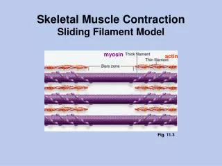
Sliding Filament. Thick & thin filaments. Myosin tails aligned together & heads pointed away from center of sarcomere. sarcomere. sarcomere. Interaction of thick & thin filaments. Cross bridges connections formed between myosin heads (thick filaments) & actin (thin filaments)

E N D
Thick & thin filaments • Myosin tails aligned together & heads pointed away from center of sarcomere
sarcomere sarcomere Interaction of thick & thin filaments • Cross bridges • connections formed between myosin heads (thick filaments) & actin (thin filaments) • cause the muscle to shorten (contract)
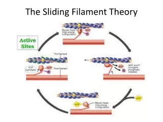
formcrossbridge releasecrossbridge shortensarcomere Where is ATP needed? binding site CleavingATP ADP allows myosin head to bind to actin filament thin filament(actin) myosin head ADP thick filament(myosin) 1 2 ATP So that’s where those10,000,000 ATPs go! Well, not all of it! 1 1 3 1 1 4
Closer look at muscle cell Sarcoplasmicreticulum Transverse tubules(T-tubules) Mitochondrion multi-nucleated
Muscle cell organelles Ca2+ ATPase of SR • Sarcoplasm • muscle cell cytoplasm • contains many mitochondria • Sarcoplasmic reticulum (SR) • organelle similar to ER • network of tubes • stores Ca2+ • Ca2+ released from SR through channels • Ca2+ restored to SR by Ca2+ pumps • pump Ca2+ from cytosol • pumps use ATP There’sthe restof theATPs! But whatdoes theCa2+ do? ATP
Muscle at rest • Interacting proteins • at rest, troponin molecules hold tropomyosin fibers so that they cover the myosin-binding sites on actin • troponin has Ca2+ binding sites
The Trigger: motor neurons • Motor neuron triggers muscle contraction • release acetylcholine (Ach) neurotransmitter
Nerve trigger of muscle action • Nerve signal travels down T-tubule • stimulates sarcoplasmic reticulum (SR) of muscle cell to release stored Ca2+ • flooding muscle fibers with Ca2+
Ca2+ triggers muscle action • At rest, tropomyosin blocks myosin-binding sites on actin • secured by troponin • Ca2+ binds to troponin • shape changecauses movement of troponin • releasing tropomyosin • exposes myosin-binding sites on actin
Coupling Excitation to Contraction • Calcium ions (Ca2+) link action potentials to contraction. • At rest, Ca2+ is stored in the sarcoplasmic reticulum. • Spaced along the plasma membrane (sarcolemma) of the muscle fiber are inpocketings of the membrane that form tubules of the "T system". These tubules plunge repeatedly into the interior of the fiber. • The tubules of the T system terminate near the calcium-filled sacs of the sarcoplasmic reticulum. • Each action potential created at the neuromuscular junction sweeps quickly along the sarcolemma and is carried into the T system.
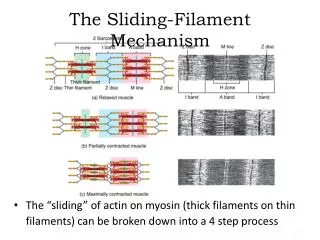
How Ca2+ controls muscle • Sliding filament model • exposed actin binds to myosin • fibers slide past each other • ratchet system • shorten muscle cell • muscle contraction • muscle doesn’t relax until Ca2+ is pumped back into SR • requires ATP ATP ATP
Thick filament Thinfilaments Thin filament Myosin head (low-energy configuration ATP Fig. 50-27-4 ATP Thickfilament Myosin binding sites Thin filament movestoward center of sarcomere. Actin ADP Myosin head (low-energy configuration Myosin head (high-energy configuration P i ADP ADP + P i Cross-bridge P i
Role of Ca and Regulatory Proteins Ca2+-binding sites Tropomyosin Actin Troponin complex Fig. 50-28 (a) Myosin-binding sites blocked Ca low concentration: binding sites are covered and contraction stops Ca hi concentration: muscle contracts Ca2+ Myosin-binding site (b) Myosin-binding sites exposed
Put it all together… 1 2 3 ATP 7 4 6 ATP 5
How it all works… • Action potential causes Ca2+ release from SR • Ca2+ binds to troponin • Troponin moves tropomyosin uncovering myosin binding site on actin • Myosin binds actin • uses ATP to "ratchet" each time • releases, "unratchets" & binds to next actin • Myosin pulls actin chain along • Sarcomere shortens • Z discs move closer together • Whole fiber shortens contraction! • Ca2+ pumps restore Ca2+ to SR relaxation! • pumps use ATP ATP ATP
Sarcomere 0.5 µm M Z Z Fig. 50-26 Relaxedmuscle Contractingmuscle Fully contractedmuscle ContractedSarcomere