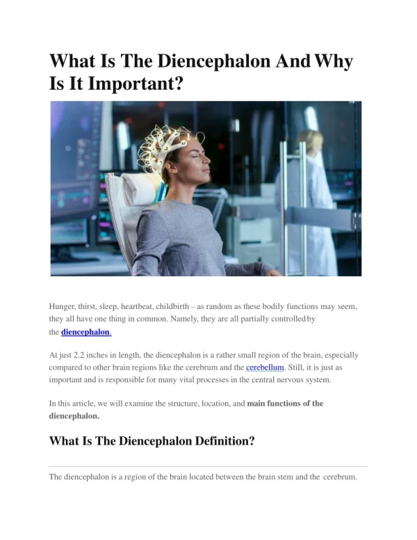
Download
1 / 24
440 likes | 1.56k Views
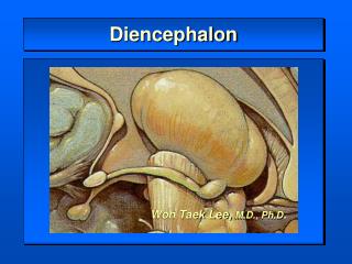
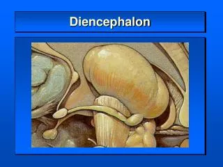
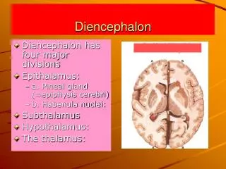
The Diencephalon. SHANDONG UNIVERSITY Liu Zhiyu. Position of Diencephalon. Position : Lies between midbrain and cerebrum, almost entirely surrounded by cerebral hemisphere. Diencephalon. Subdivision of Diencephalon. Doral thalamus Metathalamus Epithalamus Subthalamus

E N D
The Diencephalon SHANDONG UNIVERSITY Liu Zhiyu
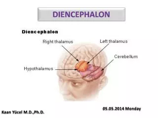
Position of Diencephalon • Position: Lies between midbrain and cerebrum, almost entirely surrounded by cerebral hemisphere Diencephalon
Subdivision of Diencephalon • Doral thalamus • Metathalamus • Epithalamus • Subthalamus • Hypothalamus
Dorsal Thalamus External features • A large egg-shaped nucleus mass • Anterior end -anterior thalamic tubercle • Posterior end - pulvinar • Right and left portion of thalamus are joined by interthalamic adhesion • Floor-hypothalamic sulcus
Classification of Nuclei of Dorsal Thalamus Three nuclear group-divided by internal medullary lamina • Anterior nuclear group • Medial nuclear group • Lateral nuclear group
Internal medullary lamina Med. nuclear group Ant. nuclear group Lateral nuclear group Medial geniculate body (MGN) Ventral anterior nucleus Ventral lateral nucleus Ventral posterior nucleus (VP) Lateralgeniculate body (LGN) Ventral posterolateral (VPL) Ventral posteromedial (VPM )
Functional Subdivision of Dorsal Thalamus Nonspecific relay nuclei-receive afferents from rhinencephalon and reticular formation of brain stem, project mainly to hypothalamus and corpus striatum • Midline nucleus group • Intralaminar nuclear group • Thalamic reticular nucleus Association nuclei-receive input from many converging sours and in turn project widely to the association areas of cerebral cortex • Anterior nuclear group • Medial nuclear group • Dorsal tier of lateral nuclear group
Functional Subdivision of Dorsal Thalamus Special relay nuclei • Vent. anterior nucleus (VA) • Vent. intermediate nucleus (VI) Receiving dentate nucleus, globus pallidus and substantia nigra to motor cortex • Vent. posteromedial nucleus(VPM ) ★ Receives trigeminal lemniscus and taste fibers • Vent. posterolateral nucleus(VPL ) ★ Receives medial lemniscus and spinal lemniscus Projects to first somatic sensory area via central thalamic radiation
Metathalamus Lateralgeniculate body (LGN) Medial geniculate body (MGN)
Metathalamus • Medial geniculate body (MGN) ★ • Relay station of audition • Receive fibers from inferior colliculus • Projects to auditory area via acoustic radiation • Lateral geniculate body (LGN)★ • Relay station of vision • Receive fibers from optic tract • Projects to visual area via optic radiation
Epithalamus Consist of: • Thalamic medullary stria • Habenular trigone • Habenular commissure • Pineal body • posterior commissure
Subthalamus • Position: transition zone between diencephalons and tegmentum of midbrain • Content:subthalamic nucleus, parts of red nucleus and substantia nigra
Hypothalamus Position-lies ventral to thalamus Boundaries • Superiorly: hypothalamic sulcus • Inferiorly: • optic chiasma • tuber cinereum • Infundibulum • mamillary body • Anterior: lamina terminalis • Posterior: continues with midbrain tegmentum
Hypothalamus Subdivisions • Preoptic region • Supraoptic region • Tuberal region • Mamillary region
Important Nuclei of Hypothalamus • Supraoptic region • Supraoptic nucleus-produce antidiuretic hormone (ADH, vasopressin) • Paraventricular nucleus-produce oxytocin Tuberal region Infundibular nucleus • Ventromedial nucleus • Dorsomedial nucleus • Mamillary region • Mamillary nucleus • Posterior hypothalamic nucleus
Paraventricular nucleus Paraventriculohypophyeal tract Supraoptic nucleus Supraopticohypophyseal tract infundibulum anterior lobe of hypophsis posterior lobe of hypophysis
Connections of Hypothalamus • Supraoptic nucleus → antidiuretic hormone (ADH) →supraopticohypophyseal tract →posterior lobe of hypophysis • Paraventricular nucleus→produce oxytocin (oxytocin) →paraventriculohypophyseal tract→posterior lobe of hypophysis • Parvicellular neurons in the arcuate nucleus and nearby region of the walls of the third ventricle secrete releasing and inhibiting hormones → tuberoinfundibular tract →portal vein of hypophysis → anterior lobe of hypophysis
Paraventricular nucleus Paraventriculohypophyseal tract Supraoptic nucleus Supraopticohypophyseal trac Inferior hypophyseal a. posterior lobe of hypophysis Hypophyseal v.
Parvicellular neurons in the arcuate nucleus and nearby region of the walls of the third ventricle secrete releasing and inhibiting hormones → tuberoinfundibular tract →portal vein of hypophsis → anterior lobe of hypophsis Tuberoinfundibular tract Median eminence Portal v. Superior hypophyseal a. anterior lobe Hypophyseal v.
Connections of Hypothalamus • Connects with limbic system • Connects with brainstem and spinal cord • Connects with dorsal thalamus • Connects with hypophysis
Functions of Hypothalamus • Autonomic control • Endocrine control • Temperature regulation • Regulation of food and water intake • Emotion and behavior • Control of circadian rhythms
Third ventricle • Position: a narrow ventricle cleft lies within diencephalons • Boundaries • Roof: choroids plexus • Floor: • optic chiasma • tuber cinereum • infundibulum and mamillary body • Anterior: lamina terminalis • Posterior: continuous with mesencephalic aqueduct • Lateral wall: dorsal thalamus and hypothalamus • Communication Third ventricle →mesencephalic aqueduct → fourth ventricle