Download
1 / 34
460 likes | 1.53k Views
Wound Healing. M . Alhashash. Definition . A wound is defined as a disruption in the normal anatomic structure and function. Wounds.

E N D
Wound Healing M . Alhashash
Definition • A wound is defined as a disruption in the normal anatomic structure and function.
Wounds • Wounds and their management are fundamental to the practice of surgery. Any elective surgical intervention will result in a wound in order to gain access to and deal with the underlying pathology. • In the surgery of trauma the wound is the primary pathology. • In both situations the surgeon’s task is • to minimize the adverse effects of the wound, • To remove or repair damaged structures , • To harness the processes of wound healing to restore function.
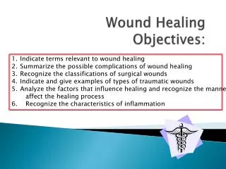
Wound Healing PHASES OF WOUND HEALING ; The wound healing process follows a predictable pattern that can be divided into : 1- hemostasis and inflammation, 2- proliferation 3- maturation 4- remodeling. This sequence of events is fluid and overlapping. All wounds need to progress through this series of cellular and biochemical events that characterizes the phases of healing to successfully re-establish tissue integrity.
1- Hemostasis and Inflammation Hemostasis precedes and initiates inflammation with the release of chemotactic factors from the wound site. Wounding disrupts tissue integrity, leading to division of blood vessels and direct exposure of extracellular matrix to platelets. Exposure of subendothelial collagen to platelets results in platelet aggregation, degranulation, and activation of the coagulation cascade resulting In a fibrin clot. Platelet granules release a number of wound-active substances such as • platelet-derived growth factor (PDGF), • platelet-activating factor (PAF), • fibronectin, • serotonin. In addition to achieving hemostasis, the fibrin clot serves as scaffolding for the migration of inflammatory cells into the wound such as polymorphonuclear leukocytes (PMNs,neutrophils) and monocytes.
1- Hemostasis and Inflammation Cellular infiltration after injury follows a characteristic, predetermined sequence. • PMNs are the first infiltrating cells to enter the wound site, peaking at 24-48h. • Increased vascular permeability, • local prostaglandin release, and • the Presence of chemotactic substances such as complement factors, interleukin-1 (IL-1), tumor necrosis factor (TNF), platelet factor 4, or bacterial products. all stimulate neutrophil migration.
1- Hemostasis and Inflammation 2. The second population of inflammatory cells that invades the wound consists of macrophages. • Derived from circulating monocytes, macrophages • achieve significant numbers in the wound by 48–96 h post-injury • and remain present until wound healing is complete
1- Hemostasis and Inflammation Macrophages, • like neutrophils ,participate in wound debridement via • phagocytosis and • contribute to microbial stasis via oxygen radical and nitric oxide synthesis. • The macrophage’s most pivotal function is activation and recruitment of other cells via mediators such as cytokines and growth factors. By releasing such mediators as TGF?, vascular endothelial growth factor (VEGF), insulin-like growth factor (IGF), epithelial growth factor (EGF), and lactate, macrophages regulate cell proliferation, matrix synthesis, and angiogenesis.Macrophages • Also play a significant role in regulating angiogenesis and matrix deposition and remodeling.
1- Hemostasis and Inflammation • T-lymphocytes comprise an other population of inflammatory / immune cells that routinely invades the wound. • Less numerous than macrophages, • T- Lymphocyte numbers peak at about 1 week post-injury • and bridge the transition from the inflammatory to the proliferative phase of healing.
2- Proliferation : The proliferative phase roughly spans days 4 through 12. During this phase, Tissue continuity is re-established. Fibroblasts and endothelial cells are the last Cell populations to infiltrate the healing wound, and the strongest chemotactic factor for fibroblasts is platelet-derived growth factor (PDGF), On entering the wound environment, recruited fibroblasts first need to proliferate, and then become activated, to carry out their primary function of matrix synthesis and remodeling. This activation is mediated mainly by the cytokines and growth factors released from wound macrophages.
2- Proliferation : Endothelial cells also proliferate extensively during this phase of healing. These cells participate in the formation of new capillaries (angiogenesis). Endothelial cells migrate from intact venulesclose to the wound. Their migration, replication, and new capillary tubule formation are under the influence of cytokines and growth factors as TNF and VEGF.
2- Proliferation : Matrix Synthesis : Collagen is the most abundant protein in the body. Type I collagen is the major component of extracellular matrix in skin. Type III, which also normally is present in skin, becomes more prominent and important during the repair process. It requires oxygen and iron as cofactors, ketoglutarate as co-substrate, and ascorbic acid (vitamin C) as an electron donor.
3- Maturation and 4- Remodeling The maturation and remodeling of the scar begins during the fibroplastic phase, and is characterized by a reorganization of previously synthesized collagen. Collagen is broken down by matrix metallo-proteinases (MMPs). The net wound collagen content is the result of a balance between collagenolysis and collagen synthesis. This will shift toward collagen synthesis and eventually establishment of extracellular matrix composed of a relatively acellular collagen-rich scar.
3- Maturation and 4- Remodeling Wound strength and mechanical integrity in the fresh wound are determined by both the quantity and quality of the newly deposited collagen. By several weeks post-injury the amount of collagen in the wound reaches aplateau, but the tensile strength continues to increase for several more months. Fibril formation and fibril cross-linking result in decreased collagen solubility, increased strength, and increased resistance to enzymatic degradation of the collagen matrix. Scar remodeling continues for many(6–12)months post-injury, gradually result in a mature, avascular, and acellular scar. The mechanical strength of the scar never achieves that of the uninjured tissue.
3- Maturation and 4- Remodeling • Epithelialization Although tissue integrity and strength are being re-established, the external barrier must also be restored. This process, beginning within 1 day of the injury, is characterized primarily by proliferation and migration of epithelial cells adjacent to the wound
3- Maturation and 4- Remodeling Re-epithelialization is complete in less than 48 h in the case of approximated Incised wounds, but may take substantially longer in the case of larger wounds in which there is a significant epidermal / dermal defect. If only the epithelium and superficial dermis are damaged, such as occurs in split-thickness skin graft (STSG) donor sites or in superficial second – degree burns ,then repair consists primarily of re-epithelialization with minimal or no fibroplasia and granulation tissue formation.
3- Maturation and 4- Remodeling Wound Contraction : All wounds undergo some degree of contraction. For wounds that do not have surgically approximated edges, the area of the wound will be decreased by this action (healing by secondary intention) . The myofibroblast has been postulated as being the major cell responsible for contraction, and it differs from the normal fibroblast in that it possesses a cytoskeletal structure.
3- Maturation and 4- Remodeling Typically this cell contains smooth muscle actin in thick bundles called stress fibers, giving myofibroblasts contractile capability. The smooth muscle actin is un-detectable until day 6, and then is increasingly expressed for the next 15 days of wound healing. After 4 weeks this expression fades and the cells are believed to undergo apoptosis. The undifferentiated fibroblasts may also contribute to wound contraction.
CLASSIFICATION OF WOUNDS Wounds are classified as either acute or chronic. Acute wounds heal in a predictable manner and timeframe. The process occurs with few, if any, complications, and the end result is a well-healed wound. Surgical wounds can heal in several ways. An incised wound that is clean and sutured closed is said to heal by primary intention . Often, because of bacterial contamination or tissue loss, a wound will be left open to heal by granulation tissue formation and contraction; this constitutes healing by secondary intention. Delayed primary closure, or healing by tertiary intention, represents a combination of the first two, consisting of the placement of sutures, allowing the wound to stay open for a few days, and the subsequent closure of the sutures. .
Classification of wound A wound can be caused by almost any injurious agent and can involve almost any tissue or structure. The most useful classification of wounds from a practical point of view is that of Rank and Wakefield into tidy and untidy wounds.
Classification of wound 1- Tidy wounds Tidy wounds are inflicted by sharp instruments and contain no devitalised tissue ; such wounds can be closed primarily with the expectation of quiet primary healing. Examples are surgical incisions, cuts from glass and knife wounds. Skin wounds will usually be single and clean cut. Tendons, arteries and nerves will commonly be injured in tidy wounds, but repair of these structures is usually possible Fractures are uncommon in tidy wounds.
Classification of wound 2- Untidy wounds Untidy wounds result from crushing, tearing, avulsion, vascular injury or burns, and contain devitalized tissue . Skin wounds will often be multiple and irregular. Tendons, arteries and nerves may be exposed, and might be injured in continuity, but will usually not be divided. Fractures are common and may be multi-fragmentary.
Classification of wound • 2- Untidy wounds • Such wounds must not be closed primarily; • If they are closed: wound healing is unlikely to occur without complications. • At best there may be wound dehiscence, infection and delayed healing, at worst gas gangrene and death may result. • The correct management of untidy wounds is wound excision, this means excision of all devitalized tissue to create a tidy wound. Once the untidy wound has been converted to a tidy wound by the process of wound excision it can be safely closed (or allowed to heal by second intention).
Factors Affecting Wound Healing • Systemic • Age • Nutrition • Trauma • Metabolic diseases • Immunosuppression • Connective tissue disorders • Smoking
Factors Affecting Wound Healing • Local • Mechanical injury • Infection • Edema • Ischemia//necrotic tissue • Topical agents • Ionizing radiation • Low oxygen tension • Foreign bodies
Treatment of Wounds • Local Care. • Antibiotics. • Dressings. • Skin Replacements. • Growth Factor Therapy.
Sutures Suture materials are divided to : 1-absorbable & 2- non-absorbable sutures:
Sutures 1- absorbable sutures : A- Plain catgut sutures : which are made from the sub mucosa of the cat intestine ,& usually absorbed in 1-2 weeks . B- Chromic catgut: It is a plain catgut but covered with chrome. It is absorbed in 3-4 weeks
Sutures C- Vicryl sutures :which are made of polyglactinic acid & absorbed in 2-3 months. D- Dexon sutures which absorbed in7-9 months.
Sutures 2- non-absorbable sutures : A- Silk :made of silky material that not absorbed. B- Nylon sutures C- Stainless steel sutures D- Cotton tape sutures
Sutures According to the type of needle: 1- cutting needle . 2- round needle . 3- taper cut needle. All these are curved needles & also there are strait needles differ in its length & thickness No needle sutures :which are used for ligation . The suture thread length about 75 cm & its thickness measured by numbers (from thick to thin) ,e.g. 2 , 1 , 0 , 2/0 , 3/0 , 4/0 up to 10/0 which is very thin suture used for eye operations .