Download
1 / 33
330 likes | 522 Views
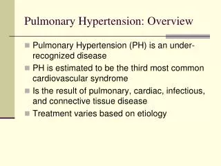
Pulmonary Hypertension. Kruti Jambula 4.15.11. Definition. PH when a disease elevates pulmonary arterial pressure above normal. Pulmonary arterial pressure = LA pressure+ (Pulmonary blood flow x Pulmonary Vascular Resistance) PA pressure >25mm Hg is abnormal.

E N D
Pulmonary Hypertension • Kruti Jambula • 4.15.11
Definition • PH when a disease elevates pulmonary arterial pressure above normal. • Pulmonary arterial pressure = LA pressure+ (Pulmonary blood flow x Pulmonary Vascular Resistance) • PA pressure >25mm Hg is abnormal. • Often progressive, if untreated RV dysfunction. • Prognosis depends on reversibility of underlying disorder.
Classification • WHO classification, initially proposed in 1998 and revised in 2003 categorizes based on pathophysiology, clinical presentation and treatment.
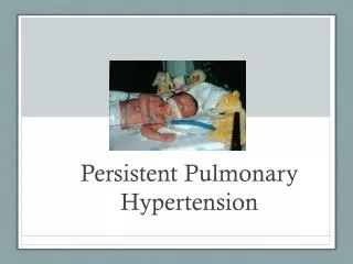
PPHN • Most common cause in newborns. • 0.2% of all term infants. • With or without acute respiratory conditions -> persistently elevated pulmonary vascular resistance -> R to L shunting across Foramen ovale/DA -> significant hypoxemia.
In the fetus systemic and pulmonary arterial pressures are almost equal because of a highly vascular placenta for gas exchange and constricted pulmonary vessels. • At birth PAP decreases to 50% systemic and PBF increases by almost tenfold. • Decline in pulmonary vascular resistance greatest in first 24 hours and continues to fall in the first 2 postnatal weeks.
Multifactorial increase in PBF • Increased arterial PH and oxygen tension • physical pulling open of capillaries accompanying lung inflation • local endogenous vasodilatory mediators- PGs and NO • removal of the low systemic vascular resistance placenta following clamping of UC.
Most newborns that have PPHN have maladaptation despite normal pulmonary arterial number and muscularization. • Mediated by a complex imbalance in local vasodilatory and vasoconstrictor metabolites- NO, PG, TAX, LKT, BK and inflammatory cytokines. • Chronic in utero hypoxia leading to increased medial muscle thickness, obstruction accompanying polycythemia or TAPV connection, pulmonary overflow following ductal narrowing, decrease in pulmonary arteries following pulmonary hypoplasia and CDH.
Presentation of PPHN • Profound and labile hypoxemia, out of proportion to parenchymal disease- suggestive, not diagnostic. • acutely ill in DR or gradually escalating signs - cyanosis, grunting, flaring, retractions, tachypnea, tachycardia and shock. • precordium normoactive unlike CHD. • S2 single and loud, TR systolic murmur. • BP/ perfusion- normal or cardiogenic shock. • Hypoxemia and acidosis constricts pulmonary vascular SM further- vicious cycle.
If shunting exclusively at Ductus: pre and post ductal Pao2 gradient of greater than 20mm Hg and a similar gradient in oxygen saturation with a decrease in postductal of greater than 5%. • Absent gradient because of shunting at atrial level or intermittent shunting. • XR: underlying lung disease or remarkably clear with diminished vascular markings and a slightly dilated heart- Idiopathic PPHN. • EKG- Normal
Echo: exclude cyanotic heart disease. • R to L shunting across foramen ovale or DA. • Deviation of atrial septum from R to L. • RA enlargement and TR.
Treatment • Aimed at preventing end-organ injury from hypoxia, ischemia and barotrauma. • Correct any contributing disturbances: hypoglycemia, polycythemia, hypothermia or pneumothorax. • Maintain systemic vascular resistance: colloids/crystalloids and inotropes • Lower pulmonary vascular resistance: iNO which activates soluble guanylate cyclase and increases cGMP-> activation of cascade causing calcium efflux and vascular SM relaxation.
iNO: • activates soluble guanylate cyclase and increases cGMP-> activation of cascade causing calcium efflux and vascular SM relaxation. • Rapid deactivation by reduced Hb leads to decreased systemic effects. • Improves oxygenation and reduces need for ECMO by ~40%, no decrease in mortality.
ECMO: Only the sickest patients are now referred. • Delay in starting ECMO, longer durations, increased age at initiation, increased rates of ECMO complications. • Survival varies with cause of PPHN.
Outcomes of PPHN • Depends on etiology, severity of hypoxemia and resultant encephalopathy. • Maldevelopment of pulmonary parenchyma and vasculature have worse prognosis. • Survivors have an increased incidence of neurodev. impairment, neurosensory hearing loss, behavioral problems and respiratory difficulties.
PH in Infants and Children • Most common causes are CHD and Pulmonary disease. • signs and symptoms are nonspecific, maybe overshadowed by underlying disease process. • Dyspnea on exertion and fatigue that progress because Right heart cannot increase CO. • Signs of right heart failure and Syncope on exertion. • Death from hypoperfusion of subendocardial tissue due to increased wall stress and increased myocardial demand or from compression of L MCA by an enlarged pulmonary artery.
Diagnostic studies • EKG: Evidence of RVH or Cor pulmonale. • 2D Echo with Doppler: Confirmatory test. • Tricuspid valve regurgitant velocity + RA estimated pressure = RV Pressure. • In the absence of PV stenosis this equals PA pressure. • Indicators of severity: degree of RV pressure elevation and reversibility.
Cor Pulmonale • Alteration in right ventricular structure and function due to PH caused by disease affecting the lung or its vascular bed. • Does not include left sided heart failure. • Rule out CHD or acquired left-sided heart disease prior to diagnosis. • If RVH seen on CXR or EKG - 2D Echo to diagnose.
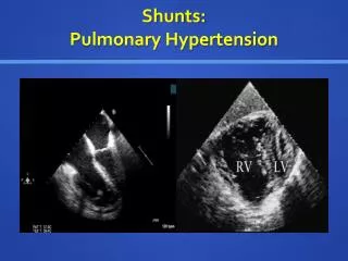
Congenital Heart disease • Pulmonary blood flow is increased, or some other factor increases downstream resistance to blood flow through lungs. (MS, PV obstruction, LV dysfunction) • PAH most common mechanism. • Significant shunt from systemic to pulmonary circuit. • Intracardiac: ASD/VSD/endocardial cushion defects • Extracardiac: PDA, Aortopulmonary window
Adapted from Nadas AS, Fyler DF. Pediatric cardiology. Philadelphia: WB Saunders, 1972:684,
L to R shunt -> Increases flow in pulmonary vascular bed -> Pulm. HTN • Shear stress on endothelial wall -> Pulm. Arteriolar endothelial dysfunction -> Arteriolar beds’ SM proliferates and hypertrophies -> PH irreversible and progressive.
Heath–Edwards classification of pulmonary vascular changes. Grade I: Medial hypertrophy. Grade II: Cellular intimal proliferation in an abnormally muscular artery. Grade III: Occlusive changes. Medium is thickened as a result of fasciculi of longitudinal muscle, and vessel is all but occluded by fibroelastic tissue. Grade IV: Dilation. Vessel is dilated, and medium is abnormally thin (arrow). Lumen is occluded by fibrous tissue. Grade V: Plexiform lesion. There is cellular intimal proliferation (arrow); clustered around are numerous thin-walled vessels that terminate as capillaries in the alveolar wall Grade VI: Acute necrotizing arteritis.
Eisenmenger Syndrome • Once pulmonary pressure exceeds the systemic pressure-> L to R shunt reverses -> Cyanosis • Supportive care only therapy.
Pulmonary Venous Hypertension • Left atrial or ventricular disease, left sided valvular disease and pulmonary venous obstruction. • LV disease -> increase LVEDP ->primary LV failure. • Uncommon. • Viral myocarditis • Medical treatment to transplant. • PV stenosis is irreversible.
Idiopathic PH • Primary Progressive elevation of PA pressure ->RV failure. • Rare with a female preponderance 1.7 : 1 • 6- 10% are familial, AD inheritance, BMPR-2 mutation • Pathogenesis: • Vasoconstriction due to an imbalance of mediators • Vascular remodeling due to proliferation of endothelial cells and vascular SM. • Thrombosis due to coagulation abnormalities.
Treat pulmonary vasculopathy and synproms of RV failure and thrombosis. • Catheterization of right heart if targeted vasodilator therapy. • Atrial septostomy-> obstructed pulmonary vascular bed bypassed-> increased CO. • i NO responders-> CCB • i NO non responders ->PG analogs • Bosentan: inhibition of potent endogenous vasoconstrictor endothelin. • Sildenafil : Breakdown of vasodilator c GMP
Respiratory disorders • Hypoxemia ->remodeling of vascular wall->increased resistance->RVH-> RHF • Alveolar hypoventilation syndromes assoc. with thoracic cage abnormalities. • OSAS, Polycythemia
Thromboembolic disorders • Obstruction of Pulm. Arteries by a venous clot.
References • Rothstein et al. Pulmonary Hypertension; Peds Rev. Vol 30, No.2 38-47. • Moss and Adams, Textbook of cardiology. • Pulmonary Arterial Hypertension: Harrison W. Farber and Joseph Loscalzo,NEJM 2004; 351:1655-1665