Download
1 / 29
290 likes | 584 Views
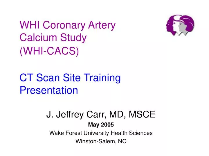
WHI Coronary Artery Calcium Study (WHI-CACS) CT Scan Site Training Presentation. J. Jeffrey Carr, MD, MSCE May 2005 Wake Forest University Health Sciences Winston-Salem, NC. Objectives. Learn about the study. Learn and understand the CT scanning protocol.

E N D
WHI Coronary Artery Calcium Study(WHI-CACS)CT Scan Site TrainingPresentation J. Jeffrey Carr, MD, MSCE May 2005 Wake Forest University Health Sciences Winston-Salem, NC
Objectives • Learn about the study. • Learn and understand the CT scanning protocol. • Opportunity ask/submit questions to the CTRC at Wake Forest!!!! • This presentation supplements the detailed material located in the 3-ring binder at each CT scan site.
What is WHI-CACS? • Women’s Health Initiative (WHI) Coronary Artery Calcium Study (full abbrev. = WHI-CACS) • Is a sub-study of the WHI involving women who received either Estrogen or placebo to better understand the role of hormone replacement. • The objective is to measure coronary atherosclerosis by CT and compare those who received estrogen to those who received placebo. • In WHI-CACS we will measure Coronary Artery Calcified Plaque (CACp) using a CT scanner with cardiac gating capability.
How is the study organized? • ~30 WHI Clinical Centers will recruit and CT scan participants. • CT Reading Center • Wake Forest U. Health SciencesWinston-Salem, NC will receive and measure the CT scans for Coronary Artery Calcified Plaque • Data Coordinating Center • Fred Hutchinson Cancer Research Center will perform analysis.
CT exams will be sent to WFU via FEDEX on CD Site 24 CT CT Reading Center will send the results to the data coordinating center WHI Coordinating Center Seattle, WA WFUCT Reading Center Site 1 CT Site 2 CT CT exams scanned at the ~30 Field Centers, ~ 900 total exams Site 25 CT
Let’s Learn the CT Protocol! • CT scanning protocol • Sending the images to Wake Forest • Quality Control (QC) procedures / Phantom CT scans
CT Visit - Overview • Greeting participant • Make sure they are the “correct” person • Female • Here for “WHI-CACS CT Scan” • Prepare participant by indicating there will be several times they are asked to hold their breath • Scout / topogram /scanogram images • Watch how they do – provide further instructions • Heart scan w/ECG • Help participant exit CT scan suite
Participant Preparation • Instruction prior to scan (clinic staff) • No caffeine products before scan (3 hrs) • Have loose clothing to facilitate placing ECG leads • No metal/jewelry around chest - this includes under-wire bras which will need to be removed or repositioned • Have clinic fax / deliver participant CT completion forms prior to study.
Entering Participant Information Into the CT Scanner • Maintain confidentiality • Positively identify each person • Identifiers have been assigned by the coordinating center – two codes are used: • Study ID number (SID) • Name code
CT data entry WHI-CACS Name: BCABCDEL ID number: 1201234L Date of Birth: 03 MAR 1947 Technologist: 51 (initials or ID) Indication: WHI-CACS
How do you know what to type in to the CT scanner? • Each participant is consented & screened by the clinic prior to the CT exam • Consent form is sign ahead of time • The participant is scheduled for the CT exam • You will be sent a WHI-CACS CT Worksheet which will have the needed information.
Participant CT Worksheet <<participant DOB>> DOB Replace “real name” Study ID
CT Worksheet Tech ID Let us know any problems! Fax back to WHI Clinic
CT Scanning overview • Scouts - Thoracic • Frontal • Lateral (optional) • Heart w/ ECG gating • Late Diastole: • EBCT 80% • GE MDCT 70% • Siemens 50% • Others TBD • We will provide a detailed protocol sheet for each specific CT scanner.
Breathing Instructions “Take a deep breath in <pause> Blow it all the way out <pause> Take a deep breath in <pause> Blow it all the way out <pause> Take a deep breath in and hold it. ******Scan 20-40 seconds***** Breathe and relax”
The Breathing Instructions are very Important! • Do the same instructions for scout and heart scans. • Why deep breath in? • Pushes liver down, • Valsalva lowers heart rate, • Blowing off C02 (hyperventilation) makes it easier to hold breath! • Use custom pre-recorded voice if available. • Have participant do the best they can – breathe out slowly if they cannot hold the entire time.
ECG – Lead placement • Arms overhead • Reposition leads if necessary • Make sure amplitude (i.e. size) is OK per your CT scanner instructions
ECG positioning • Manufacturer or local recommendations • “White is Right – Smoke (black) over Fire (red) (left side) • Must have identifiable R-wave, if not reposition & check connections • Prospective triggering/gating - late diastole phase • EBCT @ 80% (636-736) • Siemens at 50%-TBD, (428, 755) • GE at 70% ( 437-763) • Phillips/Toshiba - TBD
Check Thorax Scouts for: • Participant centered? • Prescribe cardiac scan • Carina thru inferior aspect of the heart • Lateral scout may be useful to prescribe AP centering of reconstructions • Scan using a 27 cm FOV !!!!!!!! Which should be centered on the heart - if you clip any of the heart reconstruct adjusting offsets as appropriate.
End Start Start End Scouts –Landmarks for Prescribing Scans
Cardiac Scan • Check coverage • Check centering • Check ECG • Confirm correct technique (KV, mAs, slice thickness 3 mm or 2.5 mm) • Make sure participant holds breath according to protocol
Archive & Make a CD for us • Archive the exam locally - this is to cover the possibility that some how the data is lost during the transfer • Burn the entire exam (or multiple exams) to a CD • Label the CD and place in FedEx envelope.
Sending CT scans to the Wake Forest CT Reading Center • Scans will be transferred to the CT reading via FedEx • We will provide you address labels so you can charge the costs to us • If possible send a weeks worth of scans at one time. If however no additional WHI scans are scheduled go ahead and send the CD. • Contact us with your questions • Chris O’Rourke • corourke@wfubmc.edu 336 716 8909 • Main: 336.716.7234
Scales Require Calibration - So do CT Scanners! • We all know that you can “adjust” the zero value of a scale to weight “heavy” or preferably “light” • CT Scanners can have the CT number value of water (0 H.U.) adjusted to be “brighter” or “darker”
Why are we scanning the Torso & QCT phantoms? • To make sure your CT scanner’s ability to measure CT numbers (HU) is correct • To better understand any potential differences between CT systems at the different sites
QC Scan Procedure – What Will I have to Do? • CT Reading Center will FedEx the phantoms to your site, including instructions *** Save the Box *** • Follow the instructions - The idea is to scan both phantoms with the same technical factors as we plan for the WHI participants • Call us if you have any questions - the QC scans should take less than 15 minutes to set-up and complete • Burn the exam to CD and FedEx the CD to us at Wake Forest • Re-Package the Phantoms and send to the next WHI site as instructed.
Centertorso plug Torso phantom (oval) QCT Calibration phantom (rectangle) 4 tubes containing:0, 50, 100 & 200 mg/cc Calcium QCT Phantom – PartsSetup for Quality Control scans
Cardiac Phantom Setup This should be at isocenter using your laser alignment system
Thank You! We are here to help!Please Complete your Pre-Certification Form Now • Call or Fax us with your questions! • Voice: 336.716.7234 • Fax: 336.716.4340