Download
1 / 1
10 likes | 341 Views
B. Genome-wide transcription analysis of interaction between the human macrophage and Mycobacterium tuberculosis during concurrent drug administration by conventional and novel methods. Awadh Bihari Yadav , Amit Kumar Singh and Amit Misra.

E N D
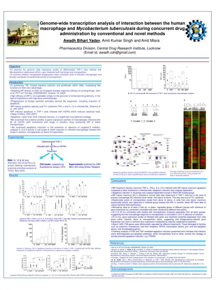
B Genome-wide transcription analysis of interaction between the human macrophage and Mycobacterium tuberculosis during concurrent drug administration by conventional and novel methods Awadh Bihari Yadav, Amit Kumar Singh and AmitMisra Pharmaceutics Division, Central Drug Research Institute,Lucknow (Email id; awadh.cdri@gmail.com) Objective • To examine the genome wide expression profile of differentiated THP-1 cells infected with Mycobacterium tuberculosis (M.tb.); upon treatment with free drugs and microparticles. • To examine whether microparticle phagocytosis alters activation state of infected macrophages and thereby contributes to bactericidal activity of microparticles.. Introduction • In pulmonary TB, inhaled bacteria colonize and proliferate within AM, modulating M functions to their own advantage. • Targeting MP directly to AM via inhalation therapy improves efficacy of existing drugs. (Sen et al,PCT Int’l Pat App. 20050084455, October 16, 2003) • High efficacy of MP is not explicable simply on the grounds of enhanced drug delivery, in the light of experience with high-dose chemotherapy. • Phagocytosis of foreign particles activates several M responses, including induction of apoptosis. • MP induce oxidative radicals and Th1 cytokines TNF- and IL-12 in infected M. (Sharma et al, JAC 2007) • MP induces apoptosis in THP-1 cells infected with H37Ra which reduces bacterial load. (Yadav & Misra, AAC 2007) • Apoptosis, rather than M.tb.-induced necrosis, is a legitimate host-defense strategy. • We examined the cytokine profiles & gene expression profiles of macrophages infected with M. tb. H37Rv after treatments with drugs in solution, drug containing MP or blank microparticles. • We examined apoptosis induction in the presence or absence of caspase-3 inhibitor, caspase-3, 8 & 9 activity in cell lysate & nitirte induction in infected macrophage treated with drugs in solution, microparticles or blank microparticles. A. CFU in cell lysate B. NO estimation in THP-1 (bar) and primary macrophages (scatter) Experimental Differentiated THP-1 Infected with H37Ra or H37Rv Treatment RNA ( 0, 12 & 24 hrs): Affymetrix HG U133 Plus 2.0 arrays; labeling, hybridization, scanning and data analysis at TCGA, New Delhi. Cell lysate: caspases by fluorescence assays; CFU. Supernatants cytokines by CBA (BD); NO using Greiss’ Reagent Two way clustering of gene & treatments at 0, 12 & 24 hrs time point , using Cluster and view in Treeview software Apoptosis pathway analysis using GenMAPP ( Free available software) at least 2 fold up or down regulated gene were considered as significant Results Conclusions • MP treatment induces maximumTNF-α , IFN-γ & IL-10 in infected cells MP induce maximum apoptosis compared to other treatments in infected cells. Apoptosis induction was caspase dependent. • Apoptosis induction in all groups was caspase dependent except in Blank MP treated group. • MP induce maximum NO in infected or normal cells after treatments in THP-1 cells but in the case of primary macrophage NO induction level varied from donor to donor. This was also true of Th1 cytokines • Bactericidal action of microparticles varied from donor to donor, in cells from one donor maximum bactericidal activity was observed in infected group treated with MP, in another, blank MP were able to reduce bacterial load significantly. • Microarray data on at least 2 fold up- or down- regulated genes of different groups with reference to gene expression by uninfected, untreated cells were clustered at different time points. • At 0 & 24 hrs uninfected cells treated with microparticles clustered separately from all other groups, suggesting that the macrophage response to microparticles is concluded in 24 h in absence of infection. • At 12 hrs, gene expression profile of infected cells given any treatment clustered separately from cells given either infection alone or microparticles alone, suggesting that phagocytosis-induced gene expression has more in common than expression modulated by infection subjected to treatment. • About 1400-1700 genes were differentially regulated by different treatments. These belonged to families such as cytokines, chemokines, and their receptors, GPCR, transcription factors, pro- and anti-apoptotic genes, and housekeeping genes. • Pathway analysis of FAS and TNF-mediated apoptosis induction presented here indicates that infection alone downregulates pro-apoptotic pathways, while microparticles tend to either reverse this strategy or actively promote apoptosis induction, e.g. TRADD. Cytokine TNF-, IFN-γ, IL-2, IL-4, IL-6 and IL-10 by THP-1 cells after infection and treatment with Rifabutine+Isoniazid; either soluble or as MP or blank MP for 2h. References • Sen et al,PCT Int’l Pat App. 20050084455, October 16, 2003) • Sharma, R., Muttil, P., Yadav, A. B., and Misra, A. Uptake of inhalable microparticles affects defense responses of macrophages infected with Mycobacterium tuberculosis H37Ra. J. Antimicrob. Chemother. 2007 59: 499-506 • Cynamon MH, Zhang Y, Harpster T, Cheng S and De Stefano MS. High-dose isoniazid therapy for isoniazid-resistant murine Mycobacterium tuberculosis infection. Antimicrob Agents Chemother 1999;43:2922-4 • Schnyder J, Baggiolini M. Role of phagocytosis in the activation of macrophages. J Exp Med 1978;148:1449-57 • A. B. Yadav and A. Misra, Enhancement of Apoptosis of THP-1 Cells Infected with Mycobacterium tuberculosis by Inhalable Microparticles and Relevance to Bactericidal Activity. Antimicrobial Agents and Chemotherapy; 2007; 51(10):3740-3742 • B. Raju,, Y. Hoshino and M. D. Weiden, Gene expression profiles of bronchoalveolar cells in pulmonary TB. Tuberculosis 2008; 88 (1): 39-51. Annexin V Staining: Fig. A. Apoptosis induction in the terms of index in THP-1 infected with H37Rv after different treatments. B. Apoptosis induction in the presence or absence of caspase-3 inhibitor. Acknowledgments Funded by the NMITLI program of CSIR. ABY is a recipient of a SRF and Amit K Singh a JRF from CSIR. We thank A.L. Vishwakarma for help with flow cytometry. Caspase Activity Assay: Specific activity of caspase- 3, -8 & -9 in cell lysate after infection with H37Rv and different treatments.