Download

1 / 66
1.04k likes | 2.54k Views
Pediatric Genitourinary Disorders. Enuresis. Repeated involuntary voiding or incontinence by a child past the age of toilet training. about 5-6 years of age. Enuresis Multitreatment Approach. Fluid Restriction Bladder exercises Timed voiding Enuresis alarms

E N D
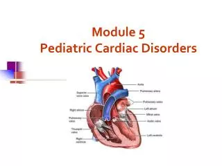
Pediatric Genitourinary Disorders
Enuresis Repeated involuntary voiding or incontinence by a child past the age of toilet training. about 5-6 years of age
EnuresisMultitreatment Approach Fluid Restriction Bladder exercises Timed voiding Enuresis alarms Reward system Medications
Urinary Tract Infection
Test Yourself • Which of the following organisms is the most common cause of UTI in children? a. staphylococcus b. klebsiella c. pseudomonas • escherichia coli All are causative agents, Escherichia coli is the more common cause of first time UTI’s.
Urinary Tract Infections • Etiology and Pathophysiology • Tend to occur more in girls than in boys because the urethra is shorter in girls and is located close to the vagina and anus. • Pathogens enter as an ascending infection • Most common causative organism is Escherichia coli
Assessment • Typical symptoms of older children and adults – dysuria, frequency, urgency, burning,hematuria – may not be present. • Symptoms not always clear • Fever • Mild abdominal pain • Bedwetting (enuresis) • If gets worse – high fever, flank pain, vomiting, malaise
Diagnostic Tests • Urine for culture and sensitivity • Clean catch • Suprapubic aspiration • Catheterization • A Positive Test • Bacteria colony count is more than 100.000/ml. • Proteinuria may also be present indicating presence of bacteria.
Therapeutic Interventions • Drug Therapy • Antibiotics – specific to causative organism • Analgesics – Tylenol • Nursing Care • Force fluids – childs choice • Dysuria – sit in warm water in bathtub and void into the water
Therapeutic Interventions • Parent Teaching • Change diaper frequently • Teach girls to wipe front to back • Discourage bubble baths • Encourage children to drink periodically during the day • Bathe daily • Adolescent start menstruating – encourage change of pad every 4 hours • When girls become sexually active – teach to urinate immediately after intercourse
Evaluation • Follow up • Return for repeat urinalysis – usually after 72 hours of treatment to be sure treatment is working • Girls who have more than three UTI’s, and boys with first UTI should be referred to urologist for further evaluation.
Common Sites for Obstruction • Stenosis of ureteropelvic valve • Stenosis of ureterovescicular junction • Stenosis of the posterior urethral valve
Pathophysiology • Reflux occurs because the valve that guards the entrance from the bladder to the ureter is defective from: • Primary reflux – congenital abnormal insertion of ureters into the bladder • Secondary reflux – repeated UTI’s cause scarring of valve • Bladder pressure that is stronger than usual, neurogenic bladder • Backflow happens at voiding when bladder contracts, urine is swept up the ureters • Results in stasis of urine in ureters or kidneys which in turn leads to infection or hydronephrosis.
Clinical Manifestations • Fever • Vomiting • Chills • Straining or crying on urination, poor urine stream, • Enuresis (bedwetting), incontinence in a toilet trained child, frequent urination. • Strong smelling urine • Abdominal or back/flank pain
Diagnostic Tests • Urine culture – done every 2-3 months • cystourethrogram • renal ultrasound - a non-invasive test in which a transducer is passed over the kidney producing sound waves which bounce off the kidney, transmitting a picture of the organ on a video screen. The test is used to determine the size and shape of the kidney, and to detect a mass, kidney stone, cyst, or other obstruction or abnormalities.
Therapeutic Interventions • Drug Therapy • Antibiotics • Penicillin • Cephalosporins • Urinary Antiseptics • Nitrofurantoin • Surgery • Repair of significant anatomical anomalies, uretheral implantation
Nursing Care following surgery • Keep accurate record of intake and output. Keep records from stents and catheter separate. Decreased output from stent could indicate obstruction. • Secure stents and catheter to prevent displacement. • Assess vital signs for signs of infection. • Assess pain. Handle child gently Administer pain medications • Patient Teaching - regarding prevention of UTI, - importance of taking all antibiotics, continue takingantiseptics even when have no symptoms.
Evaluation • Follow-up • Go in for a VCUG (voiding cystourethrogram) after a few months
Cryptorchidism Hypospadius / Epispadius Structural Defects
Cryptorchisism Failure of one or both of the testes to descend from abdominal cavity to the scrotum
Etiology and Pathophysiology • Testes usually descend into the scrotal sac during the 7-9 month gestation • They may descend anytime up to 6 weeks after birth. Rarely descend after that time. • Cause unknown • Theories • Inadequate length of spermatic vessels • Lowered testosterone levels
Why is it important that the testes are in the scrotal sac?
Answer: • The higher temperatures in the abdomen than in the scrotum results in morphologic changes to the testis – mainly concerned with lower sperm counts at sexual maturity.
Assessment Diagnosed on Newborn Physical Exam Palpate the testes separately between thumb and forefinger, with thumb and forefinger of other hand over the inguinal canal.
Therapeutic Interventions • Surgery • Orchiopexy done via laproscopy • Done around 1 year of age • Nursing Care – Post-op • Minimal activity for few day to ensure that the internal sutures remain intact • Allow opportunity to express fears about mutilation or castration by playing with puppets or dolls.
Hypospadias Epispadias
Hypospadias • Congenital urethral defect in which the uretheral opening is on the lower aspect of the penis and not on the tip.
Epispadias • Congenital urethral defect in which the uretheral opening is on the upper aspect of the penis and not on the end
Etiology and Pathophysiology • Epispadias – rare and often associated with extrophy of bladder. • Hypospadias • Occurs from incomplete development of urethra in utero. • Occurs in 1 of 100 male children. Increased risk if father or siblings have defect. • Defect ranges from mild (meatus is just below tip); to meatus on the perineum between scrotum, ventral foreskin lacking • May have accompanying chordee (a fibrous band that causes the penis to curve downward), • Undescended testes – found in conjunction with hypospadias • Might interfere with fertility in the mature male if not corrected.
Assessment Usually discovered during Newborn Physical Assessment
Ask Yourself? • Why would the nurse question an order to prepare the infant for a circumcision?
Answer: • The nurse would question the order for a circumcision because the foreskin is used in reconstruction and repair of the defect.
What is the relation of epispadius or hypospadius to infertility?
Answer: • If the urethral opening is not at the end of the penis, then the male will not be able to deposit his sperm at the opening of the os of the cervix.
Interventions • Medical Treatment: • Surgery • Reconstructive – repositions uretheral opening at tip of penis • Stent placed in urethra to maintain patency • Chordee – released and urethra lengthened.
The reason for surgery at about 1 year of age is because: a. children will experience less pain b. chordee may be reabsorbed c. the child has not developed body image and castration anxiety d. the repair is easier before toilet training C= answer
Post –op Nursing Care 1. Assess bleeding - Bleeding is controlled post-operatively by the use of pressure dressings. However, a small amount of bleeding for the first several days post-operatively is normal. A few drops of blood or a spot no larger than a quarter on the diaper is acceptable. 2. Maintain urinary drainage – care for catheter – foley / suprapubic, or urethral stent. Use double diapering.
A double diapering technique protects the urinary stent after surgery. The inner diaper collects stool and the outer diaper collects urine.
3. Control Bladder Spasms - usually due to the presence of the in-dwelling catheters are common post-operatively and are controlled by medications that relax the bladder (ie. Antispasmotics- Pro-Banthine and Ditropan) 4.Control Pain – may be given Tylenol 5. Increase fluids intake – assists in maintaining hydration and free flow of urine. 6. Do not allow to play on any straddle toys. 7. Prevent infection – no bathing or swimming until stents removed. 8. Call Dr if: • temp is over 101 • loss of appetite • pus or increased bleeding from stent • cloudy or foul smelling urine
Acute Postinfectious Glomerulonephritis
Acute PostinfectiousGlomerulonephritis Immune-complex disease which causes inflammation of the glomeruli of the kidney as a result of an infection elsewhere in the body.
Etiology and Pathophysiology • Usual organism is Group A beta-hemolytic streptococcus • Organism not found in kidney, but the antigen-antibody complexes become trapped in the membrane of the glomeruli causing inflammation, obstruction and edema in kidney • The glomeruli become inflamed and scarred, and slowly lose their ability to remove wastes and excess water from the blood to make urine.
Decreased glomerular filtration leads to accumulation of sodium and water in bloodstream causing increased intravascular and interstitial fluid volume, or edema • Protein molecules filter through the damaged glomeruli – proteinuria • Damage to glomeruli leads to hematuria. • High B/P, Heart failure may result • Common in boy 5-10 years old. Occurs 1-2 weeks after a Strep respiratory infection or after impetigo. • Has 2 phases • Edematous phase – 4-10 days • Diuresis phase
Assessment 1. Renal: a. Moderate Proteinuria b. Sudden onset of hematuria (tea-colored, reddish-brown, or smoky) and next develops oliguria c. Excessive foaming of urine 2. Cardiovascular: • a.Edema-usually eyes, hands, feet, not generalized • b.Hypertensionfrom hypervolemia which can lead to • c.Cardiac involvement CHF- orthopnea / dyspnea, • cardiac enlargement, pulmonary edema 3.Neuro a.Encephalopathy (headache, irritability, convulsions, coma-from cerebral edema)
Test Yourself • A 6 year old is admitted with R/O AGN. Which of the following symptoms would the child most likely have? a. normal blood pressure, diarrhea b. periorbital edema, grossly bloody urine c. severe, generalized edema, ascites d. severe flank pain, vomiting
Diagnostic Tests Urinalysis- protein (moderate), RBC's, WBC's, Specific Gravity elevated. *All children should have a urinalysis 2 wks after strep infection. Blood- • 1)ASO titer (antistreptolysin O) (antibody formation against Streptococcus) is elevated, indicating a recent hemolytic streptococcal infection Normal titer is 170-330 Todd units; IgG antibodies against Streptococcus may be found • 2)ESR (erythrocyte sedimentation rate) elevated showing inflammatory process • 3)BUN(urea nitrogen)& creatinine elevated indicating glomeruli damage
Therapeutic Interventions 1. Depends on the severity of the disease. No specific treatment. Bedrest encouraged. Disease is self-limiting! 2. Treat at home if normal BP & adequate output. 3. Must be hospitalized if: • BP increases • gross hematuria • oliguria present. • This way the child can be monitored closely and prevent complications. Rarely develops into acute renal failure