Download
1 / 53
700 likes | 2.15k Views
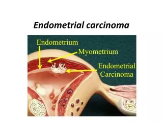
Endometrial Carcinoma and Precursors. Sigurd F. Lax. Department of Pathology, General Hospital Graz West, Graz, Austria. Endometrioid adenocarcinoma. Endometrioid histological features Well formed glands Straight luminal borders Squamous differentiation Distinct molecular genetics

E N D
Endometrial Carcinoma and Precursors Sigurd F. Lax Department of Pathology, General Hospital Graz West, Graz, Austria
Endometrioid adenocarcinoma • Endometrioid histological features • Well formed glands • Straight luminal borders • Squamous differentiation • Distinct molecular genetics • Distinct biological behavior
Endometrial hyperplasia (WHO 1994 & 2003) • Non-atypical hyperplasias • Simple hyperplasia • Complex hyperplasia without atypia (adenomatous without atypia) • Atypical hyperplasias • Simple atypical hyperplasia • Complex atypical hyperplasia (adenomatous with atypia)
Endometrial hyperplasia: Definition • Increase of glands and stroma • Simple hyperplasia: glands>stroma • Complex hyperplasia: glands>>>stroma • Simple hyperplasia: reactive due to hyperestrogenism • Atypical hyperplasia: intraepithelial neoplasia
Guidelines for Atypia Kurman RJ, Blaustein‘s Pathology of the Female Genital Tract 2002 • Atypia varies quantitatively and qualitatively • The presence of only few atypical cells does not qualify for atypical hyperplasia • ...if cellular atypia is evident without a diligent search.... • Nuclear atypia is not graded
Definition of nuclear atypia in endometrial hyperplasia (WHO 2002) • Not clearly defined • Best seen by comparison with adjacent normal glands • Frequently used criteria are: • Loss of polarity • Round vesicular nuclei • Coarse chromatin • Prominent nucleoli • Nuclear polymorphism
Caveat: Sampling Problems in Endometrial Hyperplasia • Focal nature may allow young women to maintain the uterus • Problems of possible underdiagnosing due to incomplete sampling, particularly, in scanty fragmented specimens • Hysteroscopy may assist in targeting focal lesions
Reproducibility of atypical endometrial hyperplasia (AEH)Zaino et al. Cancer 2006 • 306 cases of AEH: curettage, biopsy, pipelle • Panel of 3 pathologists; GOG Study • Agreement of at least 2 with primary diagnosis in 38% (all 3 in 15%) • Agreement for AEH worst (=0.28), best for diagnosis „normal“ and „inadequate“ • Best reproducibility of curettage cases
Causes for Poor Reproducibility of AEHZaino et al. Cancer 2006 • Specimen size • Small lesions • Technical factors (quality of sections, processing, staining) • Interpretation of criteria
Associated Endometrial Carcinoma after AEH Diagnosis in CurettageTrimble et al. Cancer 2006 • 2nd part of GOG study (Zaino et al.) • Hysterectomy within 12 weeks • 42.6% carcinomas (35% myoinvasive, most pT1b / IB) • 35% of carcinomas increased risk: G2, G3, >IA • Method: 26% D&C; 52.5% office curettage • Conclusion: high association of carcinomas
Associated Endometrial Carcinoma after AEH Diagnosis in Curettage • In 17-52% of cases in the literature (1963 – 2006) • Myometrial invasion in 2 – 39% of cases
WHO 1994: Problems and alternative approaches • Problems with this classification • Definition of atypia • Reproducibility of atypia (Kendall et al., 1998) • Reproducibility of carcinoma (Bergeron et al. 1999) • Alternative approaches and concepts • Simplified Classification for Biopsies (Bergeron et al. 1999) • Molecular and morphometric studies and the EIN concept(Mutter, Baak et al. 2000)
Reproducibility of endometrial hyperplasia and WD EC (Kendall et al. AJSP 1998) • 5 pathologists trained at the same institution • Proliferative endometrium, hyperplasias with/without atypia, well differentiated endometrioid adenocarcinoma • Good interobserver agreement for all lesions except for AEH • Best criteria for WD EC:glandular confluence, stromal alteration • Best criterion for hyperplasia: Gland crowding • Diagnoses are reproducible • Problem: criteria for atypia; agreement only on nucleoli
Alternative views on endometrial hyperplasia • EIN proposal (Endometrial Intraepithelial Neoplasia) • Simplified nomenclature proposal for biopsies (EN= Endometrioid Neoplasia) (Bergeron et al. AJSP 1999)
The importance of architectural and cytological changes • Kurman and Norris (1985): highest progression rate of complex atypical hyperplasia • Baak (1988): D-score (=VPS/standard deviation shortest nuclear axis/outer surface density of glands) • D-score <0: high risk of progression • VPS strongest predictor of progression • If VPS<55%, nuclear polymorphism especially important
EIN concept and terminology • Architecture: area of glands>stroma (VPS<55%) • Cytologic alteration: cytology differs between architecturally crowded focus and background • Size >1mm • Exclude mimics: benign conditions with overlapping criteria (basalis, secretory, polyps, repair, disruption artifact, cystic atrophy, lower uterine segment etc.) • Exclude cancer: mazelike or “ rambling“ glands, significant cribriforming, solid epithelial areas
Distinction between atypical hyperplasia and endometrioid adenocarcinoma (WHO 2002) • Stromal invasion: best evidence is provided by • Stromal disappearance between adjacent glands • Stromal desmoplastic response • Stromal foam cells beween glands: suggestive, not diagnostic • Papillary growth and extensive squamous metaplasia not accepted (occur also within metaplastic changes)
Type I (estrogen-related) Endometrial hyperplasia Younger age History of estrogen use Increased body mass index Serum estrone increased ER / PR positive (80%) Favorable prognosis Endometrioid carcinoma + var. mucinous carcinoma Dualistic Model of Endometrial CarcinomaBokhman 1983, Sherman &Kurman 1995, WHO 2002 • TypeII (not estrogen-related) • Endometrial atrophy, EIC • Higher age • No history of estrogen use • Body mass index normal • Serum estrone normal • ER / PR negative (90%) • unfavorable prognosis • Serous, clear cell carcinoma
What we need for clinical work • Type • Grade • Stage (FIGO/TNM) Curettage Postoperative Report
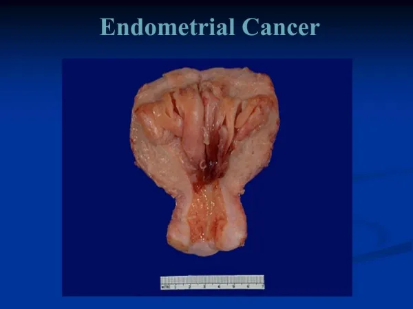
Endometrial Carcinoma (WHO 2002/03) • Endometrioid adenocarcinoma • Endometrioid adenocarcinoma- variants • Secretory endometrioid adenocarcinom • Endometrioid adenocarcinom ciliated cell type • Villoglandular endometrioid adenocarcinoma • endometrioid adenocarcinoma with squamous differentiation • Mucinous adenocarcinoma (incl. microglandular AC) • Serous adenocarcinoma • Clear cell adenocarcinoma (incl. oxyphilic type)
Endometrial carcinoma (WHO 2002/03)cont‘d • Mixed carcinoma • Undifferentiared carcinoma • Other carcinomas • Squamous cell carcinoma • Small cell neuroendocrine carcinoma • Adenoid-cystic carcinoma • Glassy cell carcinoma
FIGO Grading of Endometrioid Carcinoma • Solid, non squamous, non-morular growth pattern • 5/6-50/>50% rule • Bizarre nuclear atypia raises grade by one • Cave: Bizarre nuclear atypia should raise suspicion for serous or clear cell carcinoma
Endometrioid carcinoma with squamous differentiation • Terms adenoacanthoma/ adeno-squamous carcinoma not used • Recognition not of clinical value • Important is distinction between low grade squamous and high grade endometrioid • Grading according to the non-squamous component • Squamous component usually shows a similar grade as glandular component
Criteria for Squamous Differentiation • Keratinization (demonstrated by standard techniques) • Intercellular bridges • And/or 3 or more of the 4 following criteria • Sheet-like growth without gland formation or palisading • Sharp cell margins • Eosinophilic or glassy cytoplasm • Decreased nuclear/cytoplasmic ratio compared with foci elsewhere in the same tumor
Serous carcinoma: frequently high stageGoff et al. Gynecol Oncol 1994 • 50 patients with endometrial serous carcinoma, all surgically staged • Extrauterine disease in 72% • Lymph node metastases in 36% • No correlation with myometrial invasion and LVSI Complete surgical staging of endometrial serous carcinoma necessary regardless of depth of myometrial invasion
Clear cell carcinoma (Abeler VM, Cancer 1996) • 3% of all endometrial carcinomas • Median age 66 years • 5- and 10-year survival 42% and 39%, respectively • Relapse in 50%; 2/3 of relapse outside the pelvis • 87% diagnosed in D&C
Similarities of serous and clear cell carcinoma • Infrequent • Diagnosed at high stage • Frequent relapse • Poor prognosis • Higher age (>65) • Associated with atrophic or inactive endometrium • Low ER and PR expression; high Ki-67 labeling index
Differences between serous and clear cell carcinomas • Serous carcinoma tends to peritoneal spread • Serous carcinoma tends to be multifocal • P53 alteration seems to occur less frequent in clear cell carcinoma
Dedifferentiated endometrioid ca (Silva et al. IJGP 2006) • Undifferentiated carcinoma associated with Grade 1 or 2 endometrioid carcinoma (admixed) • 25 cases, in 50% ovaries involved • Aggressive behavior: Prognosis worse than for Grade 3 endometrioid carcinoma • Focal keratinization (9); rhabdoid features in myxoid background (6); focal neuroendocrine differentiation (4) • DD Grade 3 EC: no structure; 2 components admixed
Progression Model for Malignant Tumorsaccording to Kinzler & Vogelstein, Cell 1996 Normal Precursor Low Grade Precursor High Grade Cancer Low Grade Cancer High Grade Dysplasia Carcinoma in situ Atypical Hyperplasia Intraepithelial Neoplasia
Putative Pathogenetic Model for Endometrioid Carcinoma: “Adenoma-Carcinoma Sequence” Estrogens as driving force PTEN K-ras p53 Gatekeeper Pathway ß-catenin Normal endometrium Atypical Hyperplasia Endometrioid Carcinoma Hyperplasia MMR Deficiency/MSI Caretaker Pathway
Putative Pathogenetic Model for Serous Carcinoma: “de novo Tumorigenesis” No role of estrogens p53 p53 E-cadherin Serous intraepithelial Carcinoma Serous Carcinoma
Key genes of Type I Carcinomas • PTEN • K-Ras • Beta Catenin • Mismatch Repair Genes (MLH1, MSH2, MSH6)
PTEN • Multifunctional protein associated to the cell membrane • Induction of cell cycle arrest and apoptosis (through Akt) • signal transduction • cell adhesion and migration • neurone development • multiple other functions ??! • tumor suppressor ?! • Expression modulated by estrogens and progestin
Loss of PTEN function • Various mechanisms: • Mutation • Deletion • Promoter methylation • Downregulation? • Leads to loss of protein expression • Frequent association with microsatellite instability • Occurs in multiple tumors (breast, brain, lung, etc.)
Microsatellites • repetitive sequences with motifs up to 6 bp • > 100 000 within thegenome, usually intronic • degree of polymorphism highly variable • In part highly polymorphic within a population • replication error corrected by a particular repair system (“mismatch repair system”)
Microsatellite instability • Frameshift mutations of microsatellites • Depending on amount of altered microsatellites: MSI low (<40%) and MSI high (≥ 40%) (MSI-L/ MSI-H) • Occurs in various hereditary and sporadic neoplasms • Caused by insufficiency of the mismatch repair systems (MMR) • Mutations of repair genes (MLH1, MSH2, MSH6, PMS2) • Inactiviation of repair genes by methylation
Mismatch Repair System • Human repair system consists of dimers • Homologous to system of E. coli • Major partners: MSH2 and MLH1 • Minor partners: MSH6 (3) and PMS2 (1) • By loss of major partner minor partner unstable and dissolved
HNPCC associated tumors: Pathogenesis • Organs with increased demand of mismatch repair proteins • Environmental factors may lead to mutations or inactivation of the wild type (co-factor) • Frequently involved are colon/rectum, endometrium, stomach, small intestine, urinary tract
Familial (hereditary) Endometrial Carcinoma(Banno et al. Int J Clin Oncol 2004, Ollikainen JCO 2005, Hampel 2006) • Increased familial frequency of endometrial carcinoma with/without genetic background • Mutation-based MMR deficiency rare (2% of patients and 9% of families with familial endometrial ca) • 0.5-2% of all endometrial ca HNPCC associated • 5% of endometrial carcinomas before 55 years • Other, so far unknown mechanisms suspected
HNPCC and endometrial carcinoma Hampel et al. Cancer Res 2006 • Estimated life time risk 30-60% • Early onset (before 50 y) frequent but not exclusive • Type 1 carcinomas, low grade, low stage, favorable prognosis • 14% of surveilled mutation carriers EC in 4y (Renkonen 2006) • Not exclusively MSI-H (also MSS and MSI-L) • HNPCC criteria (Am II/Beth II) only for 30% of cases
Mismatch Repair Proteins Dimers Complex PMS 2 MSH 2 MLH 1 MSH 2 MSH6 MSH6 PMS 2 MLH 1
Repair of areplicationerror „loop formation“ MSH2 MSH6 Attachment of repair proteins PMS 2 MSH 2 MSH6 MLH 1 repair
Type II Carcinomas: P53 Mutation • Key alteration of type II endometrial carcinoma • Loss of p53 function • In > 90% of cases associated with p53 overexpression • Cave: Frameshift mutations and stop codons lead to truncated protein and a flat negative immunohistochemistry (in about 10% of mutant cases)
Wild type p53 Mutant p53 mdm 2 p21 Oncogene signals GADD DNA damage cdk Stress RB Loss of function Cell cycle arrest / G1 Bax Accumulation of mutations DNA Repair Genetic instability Apoptosis Aneuploidy
Molecular genetic differences between type I and type II endometrial carcinoma