Download
1 / 23
230 likes | 595 Views
Urinary System. The Urinary System consists of the kidneys, the ureters, the urinary bladder, and the urethra. The kidney is a bean shaped organ, reddish-brown in color, that is designed to remove waste products from the blood. They kidneys are located in the retroperitoneal space (behind the per

E N D
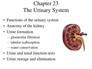
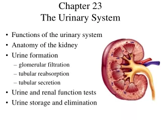
1. Chapter 20 � Urinary System
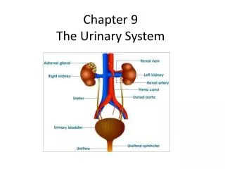
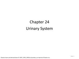
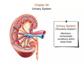
2. Urinary System The Urinary System consists of the kidneys, the ureters, the urinary bladder, and the urethra.
The kidney is a bean shaped organ, reddish-brown in color, that is designed to remove waste products from the blood. They kidneys are located in the retroperitoneal space (behind the peritoneum) in the abdominal cavity; they are affixed to the posterior abdominal wall.
3. Urinary System
4. Urinary System Structurally, the kidney has a convex surface laterally, and a concave surface medially. The concave surface has a hilus from which blood vessels, lymphatic vessels, and the ureters come and go.
The Renal Capsule is made of dense irregular connective tissue. Deep to the capsule is the Renal Cortex, and deeper yet is the Renal Medulla. The renal medulla is home to the Renal Pyramids.
5. Urinary System
6. Urinary System The tissues of the renal cortex surround and divide the renal pyramids. The renal pyramids are where the functional unit of the kidney is found. This item is called the Nephron. The nephron is responsible for maintaining blood volume, pH, and composition.
The renal arteries arise directly from the aorta and enters the kidney thru the hilum. Here they give off several branches called the interlobar arteries which pass between the renal pyramids. These then divide into arcuate arteries, that then divide into interlobar arteries. The final branches of these vessels are called Afferent Arterioles, and these lead into the nephron.
Venous blood is returned from veins in the reverse order, with the renal vein finally converging with the Inferior Vena Cava.
7. Urinary System The Nephron
Each kidney has about one million nephrons. Each nephron consists of a renal corpuscle and a renal tubule.
Each renal corpuscle consists of a tangle of capillaries called a Glomerulus, and a surrounding sac-like structure called a Bowman�s Capsule (glomerular capsule).
Afferent arterioles give rise to these capillaries which lead to Efferent Arterioles. Filtration of fluid from the glomerular capillaries is the first step in urine formation.
8. Urinary System
9. Urinary System
10. Urinary System The glomerular capsule has both a parietal and a visceral layer. The parietal layer is an expansion of the renal tubule. The visceral layer closely covers the glomerulus. The visceral layer has specialized squamous epithelium called podocytes, which in turn have specialized structures called pedicles that allow for the filtration of fluid.
The renal tubule leads away from the glomerulus and tee capsule and has three portions:
The Proximal Convoluted Tubule (PCT)
The Loop of Henle
The Distal Convoluted Tubule (DCT)
A Collecting Duct is an area where many DCT come together and drain their product (urine) into the papilla.
11. Urinary System
12. Urinary System The Juxtaglomerular Apparatus is important in the regulation of the hormone Renin. Renin is known to assist in the control of blood pressure by way of assisting Aldosterone in it�s action.
Blood entering a nephron comes in thru an efferent arteriole. The efferent arterioles have a greater diameter than the afferent arterioles. This difference in diameter causes a back up in blood in the glomerulus, leading to an increase in blood pressure in the glomerulus. This hydrostatic pressure in turn forces the filtration of blood creating a fluid called a Filtrate in the glomerular space. A part of this filtrate will later become urine.
13. Urinary System Urine Formation
Urine formation begins in the glomerular capillaries as the filtrate is formed. The substances that are allowed into the glomerular capsule via the filtrate are usually rather small in size. The pore slits between the podocytes allow for the passage of several substances, chief among which are water (of course), glucose, amino acids, urea, uric acid, creatine, creatinine, sodium, chloride, potassium, calcium, phosphate, and sulfate ions.
Many of these substances are reabsorbed later and do not end up in the final produce (urine).
14. Urinary System Large molecules, such as proteins and blood cells, are not allowed into the filtrate based on their size. They are simply too large to pass thru the slits between the podocytes. Thus, if any of these items turn up in a urine specimen a cause should be looked for as this is neither normal nor healthy.
At rest the kidneys receive about 25% of cardiac output and 20% of plasma is filtered. The filtration rate of the nephrons of both kidneys is about 125 ml per minute or 180 liters per day. However, it reabsorbs all but about 1.5 liters of this filtrate. This is a normal amount of urine for the body to create per day.
15. Urinary System Tubular reabsorption is responsible for returning water, glucose, and many electrolytes back into general circulation. The basic rules for movement of substances across a membrane must be followed in this process (i.e. diffusion, osmosis, or active transport).
Most of tubular reabsorption takes place in the PCT, but the entire tubular structure is capable of reabsorption. The peritubular capillaries are very permeable thus making the uptake of some substances even easier. Also the PCT has microvilli on some of it�s epithelia and this increase in overall surface are again helps with the reabsorption process.
16. Urinary System Glucose is generally reabsorbed in the PCT via an active process using a carrier protein. Amino acids are also reabsorbed here, these also use forms of active transport.
Water simply moves out of the tubule system by osmosis, however it is affected by the reabsorption of certain ions, and in particular sodium.
If more sodium is reabsorbed back into circulation then generally more water will be reabsorbed. This is greatly due to water being a polar molecule and sodium being a charged particle. Thus the old adage: �WATER FOLLOWS SALT.�
17. Urinary System Urea is a by product of amino acid break down in the liver. The amount of urea eliminated in the urine is a direct reflection of the amount of protein in diet. Urea is both reabsorbed and secreted, as demanded by the body. Urea in blood helps to maintain the osmotic concentration of interstitial fluid.
Uric Acid is a product of the metabolism of certain nucleic acid bases (primarily the purines known as Adenine and Guanine). Gout is an inborn error of metabolism where by uric acid crystals are deposited in joint spaces.
18. Urinary System Urine Composition
Urine is a mixture of water and solutes that are eliminated by the kidneys as a waste product. It varies mostly due to dietary intake and physical activity.
Urine is about 95% water, and will also contain urea and uric acid from the catabolism of amino acids, as well as some creatinine. Urea will also contain some amino acids, and some electrolytes.
19. Urinary System The body will usually produce from 0.6 to 2.5 liters of urine per day with an average of about 1.5 liters. This is of course subject to a variety of variable factors such as body temperature, environment temperature, humidity, respiratory rate, and some others.
Generally speaking, urine in the urinary bladder should be sterile. Not microbes should be in the bladder and presence of such is usually indicative of a bladder infection (UTI).
20. Urinary System Elimination of Urine
After forming in the nephrons, urine passes from the collecting ducts, thru the renal papilla in the pyramids, into a minor calyx. Where two or more minor calyces come together a major calyx is formed. These empty into the renal pelvis, which is a hollow area in the hilar area of the kidney.
From the renal pelvis, urine leaves the kidney via a Ureter which delivers it to the Urinary Bladder.
21. Urinary System A Ureter is a hollow tubular organ which connects the kidney(s) to the urinary bladder. The ureters have a layer of smooth muscle in their walls which is gives peristaltic contractions. These contractions help to propel the urine down and forward into the bladder.
22. Urinary System The Urinary Bladder is a hollow, distensible, muscular bag-like organ which contains urine until it is eliminated from the body. It is located in the pelvic cavity, posterior to the pubic bone on the floor of the pelvis.
The internal floor of the bladder includes an area called the urinary trigone. The urinary trigone leads to the neck of the bladder, which leads to the proximal Urethra.
The neck of the bladder forms two sphincter muscles that control the flow of urine into the urethra. The Internal Urinary Sphincter is made up of smooth muscle and is under involuntary control. The External Urinary Sphincter is made up of skeletal muscle and is under voluntary control.
23. Urinary System
24. Urinary System In a male the urethra functions both as a urinary canal and a passageway for the cells (sperm) and secretions from the reproductive organs.
Micturition is also known as urination. The micturition reflex is mediated by the sacral portion of the spinal cord. It is initiated by stretch receptors located in the wall of the urinary bladder. When the bladder fills to a certain point (between 150-300 ml) the micturition reflex will initiate and give the sensation of a need to void. The bladder usually cannot hold much more than 600 ml of urine at one time. While urinating is a voluntary action for the most part, the bladder can become overloaded at a certain point and leakage or limited flow may occur.