Download
1 / 29
300 likes | 707 Views
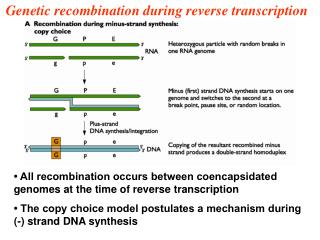
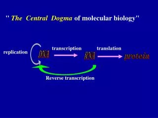
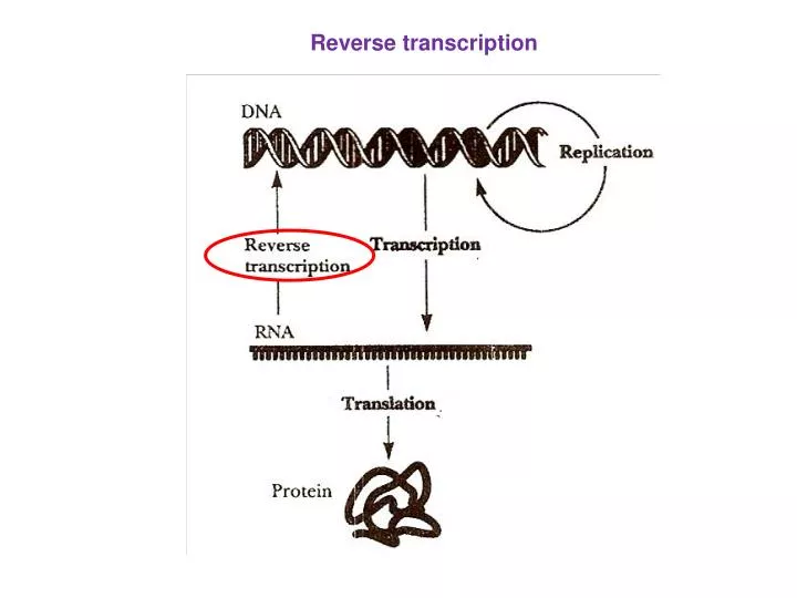
Reverse transcription. Temin ค้นพบเอนไซม์จากไวรัส (viral enzyme) ที่เปลี่ยน RNA เป็น DNA เรียกว่า RNA-directed DNA polymerase หรือ reverse transcriptase. mRNA. Reverse transcriptase. หรือ RNA-directed DNA polymerase. cDNA ( c omplementary DNA. Reverse transcriptase

E N D
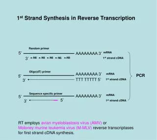
Temin ค้นพบเอนไซม์จากไวรัส (viral enzyme) ที่เปลี่ยน RNA เป็น DNA • เรียกว่า RNA-directed DNA polymerase หรือ reverse transcriptase mRNA Reverse transcriptase หรือ RNA-directed DNA polymerase cDNA (complementary DNA Reverse transcriptase - สังเคราะห์สาย DNA จากปลาย 5’ 3’ - ต้องใช้ primer(tRNA) เป็นจุดเริ่มต้นของการสังเคราะห์ cDNA
Reverse transcriptase มี 3 activities ที่ใช้ในการทำงานของไวรัส • RNA-directed DNA polymerase activity: • เติม nucleotides เพื่อใช้ในการสังเคราะห์ cDNA 2. RNase H: exonuclease สำหรับย่อย tRNA primer เมื่อมีการสังเคราะห์ cDNA แล้ว • DNA-directed DNA polymerase: • สังเคราะห์ ssDNA (single-stranded DNA)หลังจากที่ tRNA primer ถูกย่อยแล้วด้วย • Rnase Hแล้ว
Transcription mRNA capping polyadenylation nucleus Cap poly(A) Splicing Cap poly(A) cytoplasm mRNA transport Cap poly(A) Translation mRNA decay Expression of proteins from genes
Translation RNA (mature mRNA) Protein
stop start Cap poly(A) ORF 5’UTR 3’UTR mRNA structure • 5’ cap structure - 7-methyl guanosine residue • 3’ poly(A) tail - ~200 adenosine residues in mammals • ~60 in yeast • Start codon - this defines the end of the • 5’ untranslated region (5’UTR) • Stop codon - this defines the start of the • 3’untranslated region (3’UTR)
Prokaryotic ribosome Eukaryotic ribosome
Protein synthesis (Translation) • แบ่งได้ 3 stages:- • Initiation - การนำ (Recruitment) ribosome ลงบน mRNA • และจดจำ (Recognition)จุดเริ่มต้น(start codon)ของการ • translation • Elongation - การเคลื่อนที่ของ ribosome ไปบน mRNA • และแปรรหัส (Decoding) ของ mRNA และสังเคราะห์ • สายโปรตีน (polypeptide chain) • Termination - การจดจำรหัสหยุด (Recognition of the stop codon) • และปลดปล่อย ribosome และสาย protein ออกจาก • mRNA
Translation Initiation Translation initiation คือขบวนการที่ ribosome (และ initiator methionyl tRNA) ถูกนำ (recruited) ลงไปที่ start codon. เป็นขบวนการที่ซับซ้อน แบ่งได้ 4 ขั้นตอนคือ 1. การเตรียม 40S ribosomal subunit/ methionyl tRNAi • การเตรียมและการคัดเลือก mRNA • (mRNA selection and preparation) 3. การจับกันของ 40S ribosome และ mRNA, การสแกนและการจดจำ AUG (40S/ mRNA binding, scanning and AUG recognition) 4. การจับกันของ 40S และ 60S subunit (60S ribosomal subunit joining)
f-Met-tRNAfMet COOH 3’-OH : tRNA ที่จดจำ AUG (GUG, UUG, prokaryotic) 5’-P
40S/ met-tRNAi preparation 60S 60S 3 1A 40S 3 40S 1A 1 5 M GTP Ternary complex 2 M GTP M 3 2 5 1 1A GTP GDP GTP GDP 2 2 2B
mRNA selection and preparation • The mRNA is bound by eIF4F (eIF4E, eIF4G, eIF4A) • Pab1 binds the poly(A) tail and may recruit eIF4F • eIF4B and eIF4H facilitate the helicase activity of 4A
GTP 2 5 40S/ mRNA interaction eIF3 in the 40S complex and eIF4G in the mRNA complex interact 3 M 1 4G 1A 4E 4A 4B AUG Cap 4H
5 mRNA scanning • The 40S complex scans each codon in a 5’ to 3’ direction • looking for an AUG. • The eIF4A helicase activity irons out RNA hairpins • allowing the 40S complex to move. • ATP hydrolysis is required GTP 3 M 2 1 4G 1A 4E 4A 4B Cap AUG 4H
5 AUG recognition GTP 3 M 2 1 1A Cap AUG
3 1 5 GTPase step and recycling of factors • eIF5 stimulates the GTPase activity of eIF2 leading to • loss of most of the initiation factors M 1A Cap AUG GDP 2
60S Joining 60S 5B M 1A Cap AUG GTP 5B 1A GDP M Cap AUG
Prokaryotic Translation Initiation 3 1 3 fM 50S GTP 1 2 30S 30S 50S fM GTP 2 1 3 16SrRNA SHINE DELGARNO AUG
Prokaryotic Translation Initiation fM GTP 2 1 3 16SrRNA SHINE DELGARNO AUG 3 50S 1 GDP fM 2 16SrRNA SHINE DELGARNO AUG
Prokaryotic vs Eukaryotic translation • Smaller ribosomal subunits (30S and 50S) • Prokaryotic translation occurs co-transcriptionally and often there are several open reading frames in a single mRNA i.e. polycistronic mRNAs • During initiation the ribosome directly interacts with the mRNA via the Shine Delgarno sequence (directly upstream of each ORF). • Initiation is much less complicated than eukaryotes (Just three initiation factors IF1, IF2 and IF3) • Elongation and termination similar to eukaryotes
aa aa aa aa aa Translation elongation Defined as the sequential addition of amino acids to the carboxy-terminal end of the nascent peptide. Relies on three tRNA binding sites in the ribosome:- 1. The A site amino-acyl tRNA binding site 2. The P site peptidyl tRNA binding site 3. The E site where the empty tRNA is ejected from the ribosome n E P A 5’ 3’ mRNA
Four major steps:- 1. Amino acyl tRNA binding in the A site 2. GTP hydrolysis and guanine nucleotide exchange on eEF1A 3. Peptide bond formation 4. Translocation of mRNA and peptidyl-tRNA on the ribosomal surface
aa aa aa aa aa aa aa aa aa aa aa aa aa aa aa aa aa aa aa aa eEF1B Mechanism of translation elongation GDP GTP eEF1A eEF1A Amino-acyl tRNA binding n n E P A E P A 5’ 3’ 5’ 3’ mRNA mRNA Peptidyl transfer n+1 n+1 E P A E P A 5’ 3’ 5’ 3’ mRNA mRNA translocation GDP GTP eEF= eukaryotic elongation factor eEF2 eEF2
Translation termination • Catalysed by eRF1 (eukaryotic release factor). • eRF1 recognises all three stop codons and its crystal structure resembles a tRNA even though it is a protein. • Hence the molecular mimicry model predicts that eRF1 gives termination by binding the ribosome in a similar way to tRNA. • eRF3 stimulates eRF1 activity in a GTP-dependent manner